
The textbook account of mole biology says new moles appear in childhood and adolescence, stabilise by the late twenties, and rarely arise after that. Like most textbook simplifications, it's broadly true but lossy at the edges. Plenty of healthy adults develop a small number of new pigmented lesions in their thirties and forties; the question is which of those are benign acquired naevi and which warrant a consultant's eye.
The relevant variable is age. A new mole at 32 is reasonably common and usually benign; a new mole at 62 is uncommon and often deserves at least one consultant assessment. The rate of new mole formation falls steadily through adulthood, so the same lesion carries a different prior probability of significance at different decades.
This guide covers what's normal, what isn't, and the threshold for booking at each stage of adult life. Written by a GMC-registered consultant plastic surgeon for adults across the UK who've noticed something new and want a measured answer.
Mole biology, decade by decade
Mole formation peaks in childhood and adolescence, driven partly by genetics and partly by sun exposure during the period when melanocytes are most active. By the late twenties, the typical adult has reached their lifetime mole count (10-40 for most people; over 50 for some). Through the thirties and forties, the count is broadly stable, with some new moles forming and some existing ones fading or transitioning toward intradermal forms.[1]
After age 40, new mole formation slows considerably. After age 50, new pigmented lesions are uncommon enough that any individual one warrants at least a consultant's consideration, particularly if it appears on sun-exposed skin (face, neck, hands). After age 60, a genuinely new pigmented lesion should be considered de novo melanoma until proven otherwise; the threshold for booking is low.
These age thresholds are statistical, not absolute. A 65-year-old can develop a new entirely benign compound naevus, and a 35-year-old can develop a new melanoma. But the prior probability shifts meaningfully with decade, which is why the same finding triggers different urgency tiers depending on age.
What actually counts as 'new'
Patients use 'new' loosely. For clinical purposes, 'new' means a pigmented lesion that wasn't present at all six months to a year ago, not just one that's grown more visible or been recently noticed. The distinction matters because lesions that were always there but only recently noticed are usually less concerning than lesions that genuinely arose in the recent past.
Practical test. Can you find the lesion in any of: family photographs from a year ago, holiday photos showing the same body region, photos taken for unrelated reasons (e.g., posting on social media)? If yes, the lesion is older than your conscious memory of it, and you can usually relax the urgency. If no, treat it as genuinely new and apply the age-based thresholds below.
Photographs from anywhere in the past 12 months are remarkably useful for this. The casual nature is helpful: holiday snaps don't pose the body deliberately, so they often capture lesions you wouldn't have asked anyone to photograph. If you have an iPhone or Android with cloud-stored photos, scrolling back is the closest thing most adults have to a baseline mole map.
New moles in your thirties and early forties
A new mole appearing for the first time in your thirties or early forties is reasonably common. Acquired melanocytic naevi continue to form into the fourth decade, particularly in patients with significant sun exposure during young adulthood. A new lesion that looks like your other moles (similar size, shade, shape, with a regular border and uniform colour) is usually entirely benign and does not require urgent consultation.
What does warrant a consultation in this age group is a new lesion that doesn't look like your other moles, or that has any ABCDE feature on first appearance: asymmetric, multi-coloured, larger than 6mm, with an irregular border. The 'ugly duckling' principle applies; a new lesion that doesn't fit your usual mole pattern is more concerning at any age, and that includes the thirties.
Practical rule for ages 30-45. New mole that resembles your other moles, no concerning features: book a routine mole check at the next convenient time (within a few months). New mole that doesn't fit your usual pattern, or has any ABCDE feature: book within a week. New mole combined with any change in an existing mole: book within a week regardless of age.
New moles in your late forties and fifties
From the late forties through the fifties, the threshold tightens. New mole formation is still occasionally normal in this age group, but the prior probability of any specific new lesion being a problem is higher than in earlier decades. Many UK consultant dermatologists recommend that any new pigmented lesion appearing for the first time after age 50 be examined with dermatoscopy at least once.
The reasoning. After 50, the cumulative probability that an individual new lesion is a melanoma in situ or early invasive melanoma rises noticeably. Melanoma incidence in the UK peaks in the 50s and 60s.[2] So even a small lesion that looks unremarkable to the patient deserves a single consultant assessment, both to confirm benignity and to establish a documented baseline for future comparison.
Practical rule for ages 45-60. Any genuinely new mole: book within a month, even if it looks unremarkable. New mole with any ABCDE feature: book within a week. Any new lesion that itches, bleeds or crusts: book within a week.
New moles after 60
After age 60, genuinely new pigmented lesions are uncommon and warrant a consultant assessment without much further consideration. The differential diagnosis at this age includes: late-onset benign naevi (rare but real), seborrheic keratosis (extremely common harmless lesions that can look mole-like), solar lentigines (sun-spots that can resemble moles), and melanoma. Only dermatoscopy reliably distinguishes between these, and the cost of getting it wrong rises with age.
Particular caution applies to lesions on sun-exposed sites: face, scalp, ears, dorsum of hands, lower legs in women. These are the sites where lentigo maligna (a slow-growing form of melanoma in situ that prefers chronically sun-damaged skin) most often arises in older patients, and it can present as an apparently unremarkable new pigmented patch that grows slowly across years.
Practical rule for age 60+. Any new pigmented lesion: book within a month. Any new lesion on the face, scalp or sun-exposed sites: book within two weeks. Any new lesion that itches, bleeds, crusts, or looks different from your existing moles: book within a week.
A note on pregnancy and hormonal change
Pregnancy and certain hormonal events (combined oral contraceptive initiation, peri-menopause, hormone replacement therapy) can cause existing moles to darken, slightly enlarge, or become more prominent. They very rarely cause genuinely new moles to appear in adults. Hormonal changes in existing moles are not in themselves a reason for concern, but the same ABCDE thresholds apply: a mole that's changed during pregnancy in ways consistent with hormone-driven darkening (uniform, gradual, mirrored across multiple moles) is usually benign; a mole that's changed asymmetrically, developed multiple colours, or grown rapidly during pregnancy deserves a consultation regardless of the assumed hormonal cause.
Pregnancy itself does not increase melanoma risk meaningfully, but a subset of pregnant women do develop or detect their first melanoma during pregnancy partly because they're paying more attention to their bodies and partly because hormonal changes can prompt an existing lesion to evolve. Pregnant patients with concerning moles should have a consultant assessment; dermatoscopy and excision under local anaesthetic are both safe in pregnancy when needed.[3]
Practical rule. Hormonal change does not exempt a mole from ABCDE thresholds. A mole that ticks any of the warning boxes during pregnancy or hormonal transition deserves the same consultant assessment as it would outside that context. The cost of an unnecessary consultation is small; the cost of attributing a real change to hormones can be much larger.
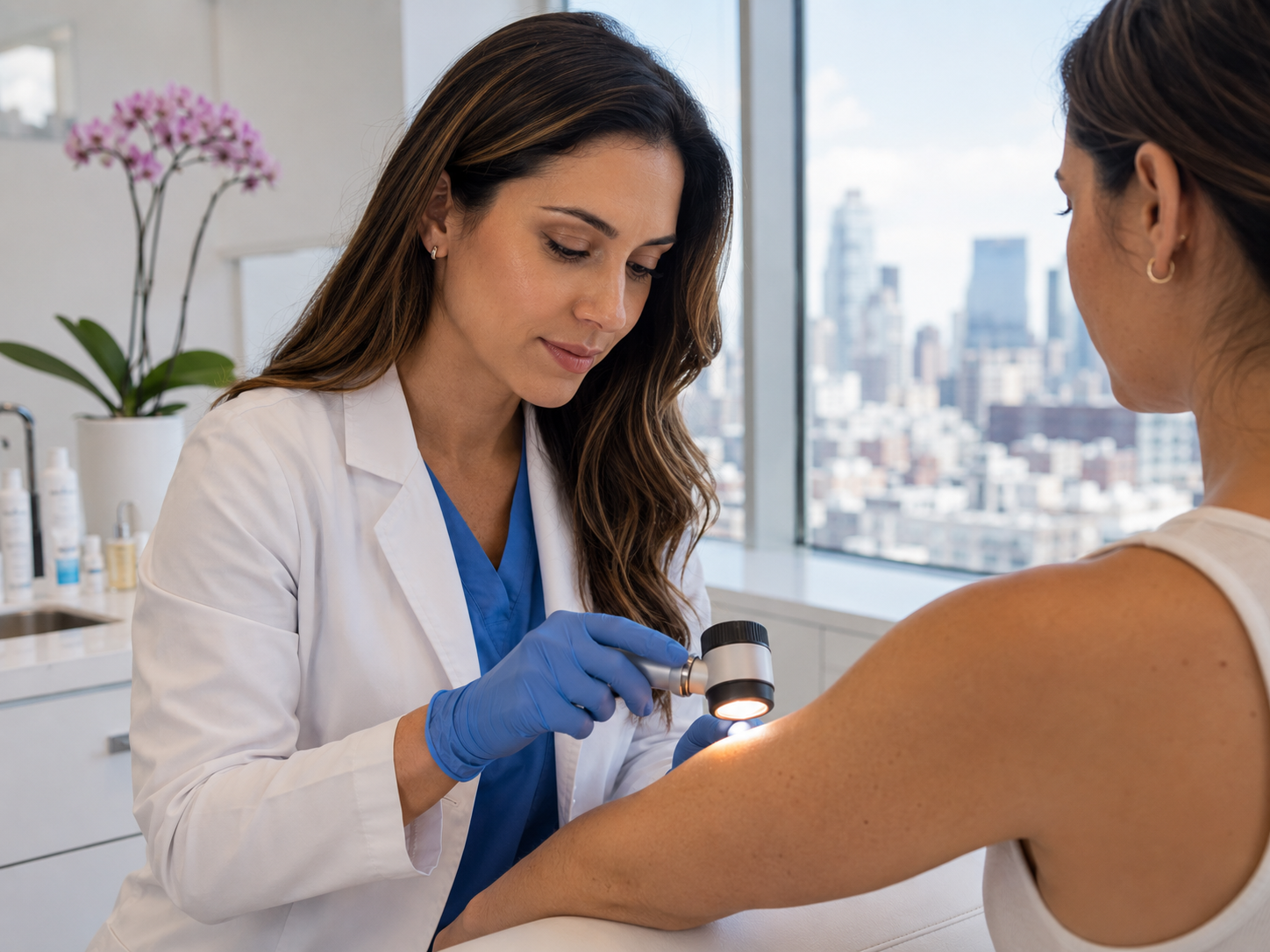
What the consultant will do
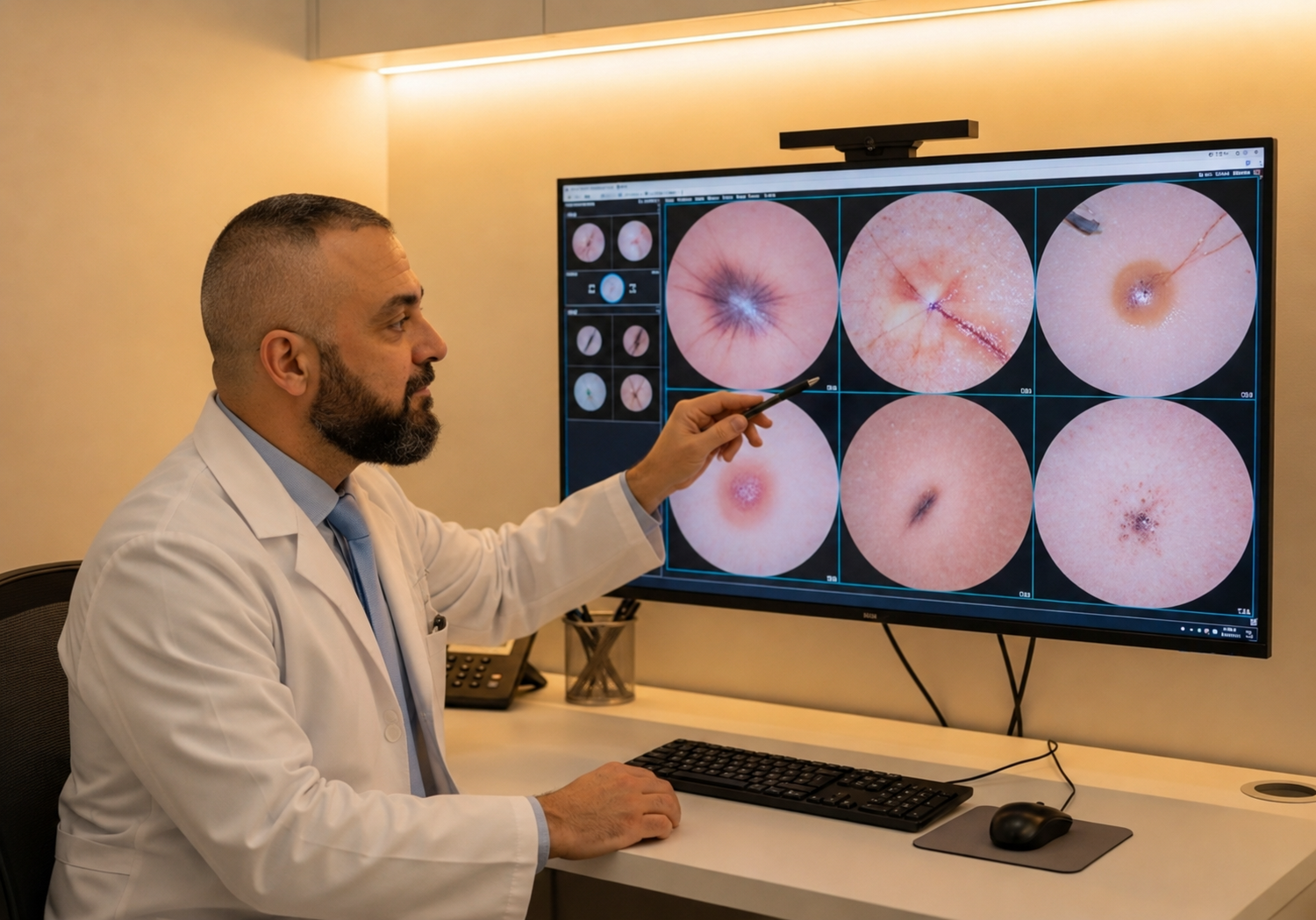
The assessment for a new mole follows the same structure as for any other concerning lesion: clinical history, naked-eye examination, dermatoscopy, plain-language discussion. Specific questions for new lesions include when you first noticed it, whether you've noticed any change since first noticing it, and whether you have any photographs that establish or refute its newness.
If dermatoscopy is reassuring, the consultant will reassure you and may recommend photographic monitoring at 3-6 months to confirm stability. If dermatoscopy is borderline, the recommendation is usually photographic monitoring at 3 months or excision with histology, depending on the dermatoscopic features. If dermatoscopy is suspicious, same-day full-thickness excision with histology is the standard recommendation.
Most patients with newly noticed moles in adulthood receive complete reassurance from a single 30-minute appointment. The minority who need treatment are usually seen, treated and reported on within two weeks of first contact. Same-week appointments, written report within 24 hours, self-pay only.
Common questions
Frequently asked
References
Sources cited
- Bauer J, Garbe C. Acquired melanocytic nevi as risk factor for melanoma development. A comprehensive review. Pigment Cell Res. 2003;16(3):297-306. View source
- Cancer Research UK. Melanoma skin cancer incidence statistics by age, UK. View source
- Pennoyer JW, Grin CM, Driscoll MS, et al. Changes in size of melanocytic nevi during pregnancy. J Am Acad Dermatol. 1997;36(3):378-382. View source
- Newton-Bishop JA, Bishop DT, Harland M, et al. Genotype/phenotype and penetrance studies in melanoma families with germline CDKN2A mutations. J Invest Dermatol. 2010;130(2):557-565. View source
- NICE guideline NG12. Suspected cancer: recognition and referral. Updated 2023. View source