
'Atypical' is one of those medical words that lands harder than it should. Hear it from a GP or dermatologist about one of your moles and the immediate fear is cancer. The clinical reality is more nuanced and a great deal more reassuring than that fear suggests, but only if you understand what the word actually means.
Atypical, sometimes called dysplastic, describes a mole whose architecture, the arrangement of melanocytes inside it, is disordered compared to a routine benign mole. It is not melanoma. The cells are not malignant. But the architecture sits on a spectrum that includes melanoma at one end, and a sizeable body of evidence shows that people with multiple atypical moles have a higher lifetime melanoma risk than the general population.
This guide explains what atypia is, why it matters, where on the spectrum your moles are likely to sit, and what the right monitoring and treatment response looks like in 2026 UK clinical practice. Written and medically reviewed by a GMC-registered consultant dermatologist.
What 'atypical' actually means
In dermatology, atypical and dysplastic are used almost interchangeably to describe a mole that, on close examination, shows architectural and cellular features intermediate between a routine benign naevus and a melanoma. The distinction is not arbitrary, it sits on a histological spectrum that pathologists have characterised in detail since the 1970s.[1]
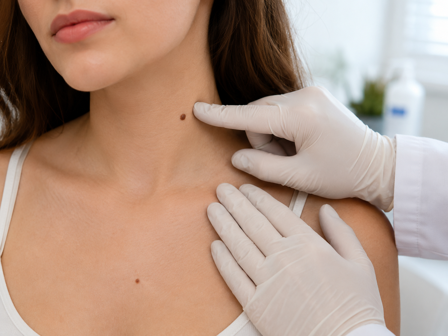
Clinically, atypical moles tend to share several visual features. They are usually larger than typical (>5mm), have an irregular or fading border, show varied colour within the same mole, and sometimes have a 'fried egg' appearance, a darker central region surrounded by a paler outer rim. Many are entirely flat. Crucially, individual atypical moles can look benign on naked-eye examination and only reveal their atypia under dermatoscopy or histology.
Importantly, 'atypical' is a clinical descriptor, not a diagnosis of cancer. An atypical mole has not become melanoma and most never will. What atypia tells us is something about the underlying biology and the patient: it is a marker of risk, both within that specific mole and elsewhere on the skin.
Atypia exists on a spectrum
Pathologists grade atypia in three rough tiers, mild, moderate and severe, each with its own management implications. Most atypical moles seen in routine practice are mild. The grading is not always cleanly reproducible (different pathologists may grade the same lesion slightly differently) but the spectrum is real and useful.[2]
Mildly atypical moles show subtle architectural disorder but otherwise look reassuring. The right response is photographic monitoring. We document the lesion, image it, and re-check at 6 to 12 months. The vast majority remain stable indefinitely.
Moderately atypical moles show clearer architectural and cytological abnormality. Annual mole mapping is the standard; some consultants will recommend excision, particularly in patients with other risk factors. The judgement call is between definite knowledge (excision plus histology) and ongoing monitoring (mapping plus re-image).
Severely atypical or severely dysplastic moles sit very close to the melanoma boundary on the histological spectrum. The right response is full-thickness excision with histology, and a wider excision if the margins are not clear. This is not the most common scenario but it is the one where the type label matters most.
Why atypia matters: the melanoma connection
The reason dermatologists pay close attention to atypia is statistical, not deterministic. People with atypical moles have a measurably higher lifetime risk of developing melanoma. The risk scales with the number of atypical moles a person has and with family history.[3]
Importantly, melanoma in patients with multiple atypical naevi is not necessarily found within an atypical mole. Plenty of melanomas in these patients arise from previously normal-looking skin or from previously typical naevi. So the right interpretation of atypia is broader than 'this specific mole might be cancer'. It is closer to 'this person's biology produces atypical melanocytes, and that has implications for how we screen them across the whole skin'.
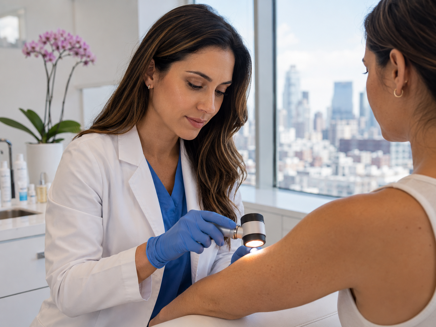
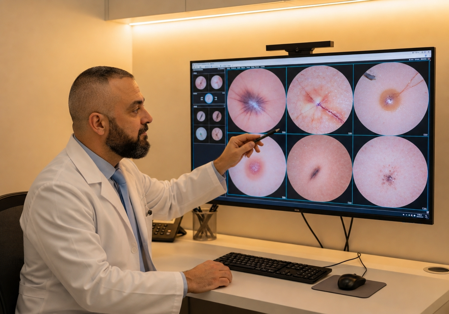
This is the foundation for the modern UK approach to atypical mole care: photographic mole mapping. Annual standardised photography of the entire skin, plus dermatoscopic close-ups of every atypical mole, gives a precise comparison year on year and catches new or changing lesions much earlier than naked-eye examination ever could.
The right monitoring strategy
For most patients with one or two mildly atypical moles, baseline mole mapping at the time of the initial diagnosis, plus an annual follow-up, is the appropriate monitoring rhythm. The 60-minute mapping appointment captures total-body photographs from set angles plus dermatoscopic close-ups of every atypical mole. At every annual visit, the previous year's images are pulled up alongside the new ones and any change is flagged immediately.
Patients with multiple atypical moles, with a personal history of melanoma, or with a strong family history may benefit from more frequent intervals (every six months for the first one to two years), particularly when establishing a baseline. Once stability is documented across two consecutive checks, most can move to annual mapping.
Between formal appointments, monthly self-examination remains valuable. The ABCDE framework is the right tool: any atypical mole that changes on asymmetry, border, colour, diameter or evolution warrants an interim consultation rather than waiting for the annual review. Patients should photograph anything they notice changing, dated phone photographs are far more useful than memory at the next appointment.
When to remove versus when to monitor
The pragmatic question every consultant faces with atypical moles is: should this come off, or should we watch it? The answer depends on three things: the degree of atypia, the patient's other risk factors, and whether there is recent change. None of those alone is decisive; together they are.
Severe dysplasia is a strong indication for excision. Moderate dysplasia in a patient with no other risk factors might reasonably be monitored; in a patient with previous melanoma it usually comes off. Mild dysplasia almost always justifies monitoring rather than excision. Anything actively changing, regardless of grade, should be excised.
When excision is recommended it is performed as a full-thickness elliptical excision under local anaesthetic in the same appointment as the consultation. The tissue is sent to a UKAS-accredited pathology lab and histology returns within 7 to 10 working days. If margins are not clear we recommend a wider local excision; if the lesion is melanoma, a multidisciplinary team meeting agrees the next step before the patient is called.
Familial atypical multiple mole melanoma (FAMM)
A small subset of patients have a strong inherited predisposition to atypical moles and melanoma, captured by the syndromic label FAMM (familial atypical multiple mole melanoma). The clinical picture: many (often more than 50) atypical moles, plus melanoma in two or more first or second-degree relatives. Some patients additionally carry mutations in CDKN2A and other melanoma-susceptibility genes.[4]
FAMM patients sit at the highest end of the atypia spectrum and benefit from intensive surveillance: mole mapping every six months, lower thresholds to excise, and in some cases genetic counselling. The condition is rare, around 1 in 10,000 in the UK, but recognising it is important because the lifetime melanoma risk is meaningfully elevated, often quoted in the 50% range over a lifetime.
If your family history includes multiple cases of melanoma in close relatives, particularly across generations, raise this with your consultant. FAMM is not a diagnosis you should self-apply, but it is one your consultant should consider and rule in or out, because the management plan looks materially different from standard atypical naevus care.
Living with atypical moles
Most patients with atypical moles live entirely normal lives, attend annual mapping appointments, and never develop melanoma. Atypia is a risk marker, not a sentence. The right mindset is informed vigilance, not anxiety.
Practical habits that make a measurable difference: SPF 30+ daily on exposed skin (rising to SPF 50+ in summer or on holiday), monthly ABCDE self-checks with a partner's eye for the back and scalp, dated phone photographs of any mole you find interesting, and a low threshold to book an interim consultation if anything changes.
What does not help: removing every atypical mole 'just in case' (impractical and unnecessary), avoiding the sun completely (vitamin D matters too), or relying on internet AI symptom-checkers as a substitute for clinical examination. The atypia label is useful precisely because it tells your dermatologist how to weight your moles. Use it that way, and let the system designed for it do the rest.
Common questions
Frequently asked
References
Sources cited
- Clark WH Jr, Reimer RR, Greene M, Ainsworth AM, Mastrangelo MJ. Origin of familial malignant melanomas from heritable melanocytic lesions. 'The B-K mole syndrome'. Arch Dermatol. 1978;114(5):732-738. View source
- Naeyaert JM, Brochez L. Clinical practice. Dysplastic nevi. N Engl J Med. 2003;349(23):2233-2240. View source
- Gandini S, Sera F, Cattaruzza MS, et al. Meta-analysis of risk factors for cutaneous melanoma: I. Common and atypical naevi. Eur J Cancer. 2005;41(1):28-44. View source
- Goldstein AM, Chan M, Harland M, et al. Features associated with germline CDKN2A mutations: a GenoMEL study of melanoma-prone families from three continents. J Med Genet. 2007;44(2):99-106. View source