
Skin cancer in darker skin types is consistently under-discussed, under-suspected, and over-represented in late-presentation statistics. Patients with skin types V and VI (deeply pigmented Black or South Asian skin) have a meaningfully lower lifetime melanoma risk than fair-skinned patients, but when melanoma does occur, it presents at later stages and on different anatomical sites, with worse outcomes.[1] The UK is no exception.
Three things drive this gap. First, the dermatology textbooks and patient-education materials that shape public awareness are overwhelmingly built around fair-skin presentations: the ABCDE framework, the typical superficial-spreading melanoma image, the sun-exposure narrative. Second, the sites where melanoma actually arises in darker skin (palms, soles, nail beds, mucous membranes) are the sites patients least often examine themselves. Third, the message 'darker skin doesn't burn easily' has been received as 'darker skin doesn't get skin cancer', which is wrong.
This guide is the version a UK consultant would write for a patient with Fitzpatrick III to VI skin who wants to understand their actual risk profile. Written by a GMC-registered consultant plastic surgeon for adults across the UK.
The real numbers
Cancer Research UK statistics show melanoma incidence in the UK is highest in the white British population, with substantially lower incidence in Asian, Black and mixed-ethnicity populations.[2] But within those groups, when melanoma is diagnosed, the survival outcomes are worse: patients with skin types V and VI present at later stages and have lower five-year survival than equivalent-stage fair-skinned patients.
Two factors drive the survival gap. Site distribution. Melanoma in darker skin disproportionately arises on acral sites (palms, soles, nail beds), mucous membranes, and rarely on chronically sun-exposed skin. These sites are under-screened both by patients (who don't routinely examine their soles or under their nails) and by clinicians (who can default to 'sun-exposed-site' assumptions).[3] Diagnostic delay. A pigmented lesion on the sole of a Black patient is more likely to be assumed to be a benign pigmented spot than the equivalent lesion in a White patient, in part because the visual pattern doesn't match the textbook. The result is later-stage diagnosis.
The clinical takeaway is not that darker skin types should worry more (in absolute terms, melanoma is uncommon at any skin type). It's that the screening framework should be matched to the actual risk profile: focus more on acral and mucosal sites, less on cumulative-UV-exposure assumptions, and build a clinical relationship with a consultant who has experience across the full Fitzpatrick range.
Acral lentiginous melanoma: the form most missed
Acral lentiginous melanoma (ALM) is a melanoma subtype that arises on hairless skin: palms, soles, fingers and toes, and nail beds (subungual melanoma). It is uncommon in fair-skinned patients but accounts for a much higher proportion of melanomas in darker skin types: around 30-50% of melanomas in Fitzpatrick V-VI patients, compared to under 5% in Fitzpatrick I-II patients.[3]
Visually, ALM presents as a slowly enlarging dark patch on a palm, sole, or under a nail. On the sole or palm, it can look like a bruise that doesn't fade, an irregular dark spot, or a slowly spreading pigmented patch with poorly defined edges. Under a nail, it presents as a longitudinal dark band (longitudinal melanonychia) that widens over months, often with extension of pigment onto the surrounding skin (Hutchinson's sign), or as a destructive lesion of the nail plate itself.
Self-check approach. Examine palms and soles monthly, ideally in good light, looking for any pigmented spot that wasn't there before or that's enlarging. Examine each nail for new dark longitudinal bands, particularly bands wider than 3mm or with irregular edges. New pigmentation extending from the nail bed onto the cuticle or surrounding skin (Hutchinson's sign) warrants a consultant within a week.
Subungual (nail) melanoma: the dark band to take seriously
Longitudinal pigmented bands in nails are common in darker skin types, with most being entirely benign (longitudinal melanonychia from physiological pigment deposition or benign nail-matrix naevi). Distinguishing benign from malignant longitudinal melanonychia is one of the harder visual judgements in dermatology, and it's an area where dermatoscopy plus expert assessment matters most.[4]
Features that warrant a consultant: a longitudinal band that's new (especially after age 40); a band that's becoming wider over months; a band with multiple shades or irregular borders; a band wider than 3mm; pigment extending from the nail bed onto the cuticle or surrounding skin (Hutchinson's sign); a single nail with a band when none of your other nails have one. Any of these features in isolation deserves an assessment within a week.
Features that are reassuring: a band that's been the same width for years; a band present on multiple nails (particularly in a consistent pattern); a uniformly shaded thin band (under 3mm) without any extension onto the surrounding skin. Even reassuring features benefit from at least one baseline consultant assessment to document the pattern, because future change is then easier to evaluate against a known starting point.
Non-melanoma skin cancer in darker skin
Basal cell carcinoma (BCC) and squamous cell carcinoma (SCC) are less common in darker skin types than in fair skin, but they do occur and they tend to present at later stages. BCC in darker skin typically appears as a pigmented (rather than the classical pearly) papule, often on sun-exposed sites; the pigmentation can make it look like a benign mole, delaying diagnosis. SCC in darker skin disproportionately arises on chronically inflamed or scarred skin (old burns, chronic ulcers, areas of long-standing dermatitis) rather than on sun-exposed skin, and tends to be more aggressive than equivalent-stage SCC in fair skin.[5]
What to watch for: any non-healing skin lesion lasting more than 4-6 weeks (regardless of pigmentation); any lesion arising on an old scar, burn site, or chronic wound that's enlarging or changing; any new pigmented papule on the face, ears, or neck that doesn't fit your usual mole pattern. Lower threshold to seek assessment on chronically scarred skin in particular.
The site distribution is the key adjustment. The traditional 'sun damage = skin cancer risk' message captures most fair-skin presentations and a smaller fraction of darker-skin presentations. Add 'chronic inflammation = skin cancer risk on darker skin' to your mental model, and the threshold to assess any persistent lesion in those areas falls appropriately.
ABCDE for darker skin: what to keep, what to add
The ABCDE framework still applies in principle. Asymmetry, border, colour, diameter and evolution remain the relevant features for cutaneous moles regardless of skin type. The adjustment is two-fold.
Pattern matching is harder. Many of the published ABCDE images and the visual training a GP receives is based on fair-skin presentations. A subtle ABCDE feature that's obvious against pale background skin can be much harder to see against deeply pigmented background skin. Patients with darker skin sometimes need to bring more aggressive change-detection (good photographs at intervals) rather than relying on a striking visual difference at a glance.
Site distribution shifts the search field. ABCDE applied to the back, chest and limbs catches most fair-skin melanomas; the same approach applied to darker skin will miss the acral and subungual presentations that account for much of the disease burden. Add palms, soles, finger and toe webs, and nails to the routine self-examination, and lower the threshold for any pigmented change at these sites.
Mole mapping for darker skin
Total-body mole mapping is genuinely useful for darker skin patients with elevated risk profiles (multiple atypical naevi, family history, immunosuppression), with one caveat: the protocol needs to include systematic coverage of acral sites and nails, which not every UK clinic does as standard.
Our mole mapping protocol includes total-body photography from set angles plus dedicated dermatoscopic close-ups of each palm, each sole, and any nail showing pigmented bands. Annual comparison year-on-year catches subtle changes earlier than naked-eye examination, which is particularly valuable for the long-tail subungual melanoma where the change is gradual across months.
Patients who benefit most: anyone with multiple atypical moles, a family history of melanoma in a first-degree relative, prior skin cancer, or immunosuppression (transplant recipients, long-term high-dose immunosuppressants). For these patients, annual mapping plus monthly self-examination of acral sites is the standard surveillance protocol our consultants recommend.
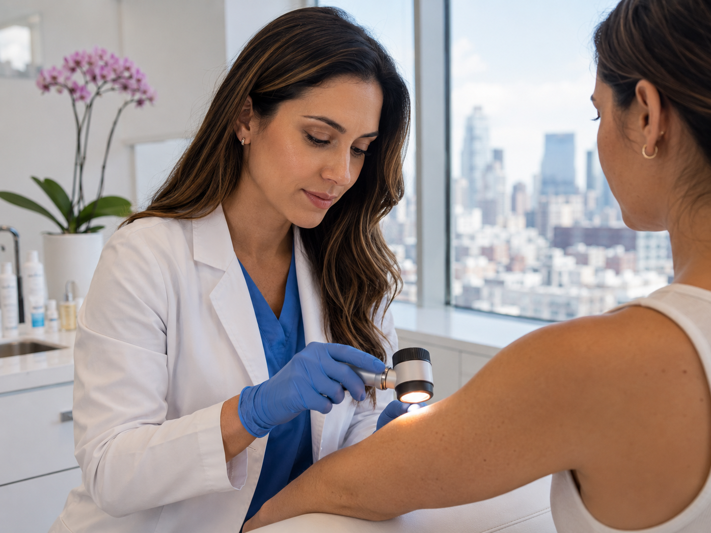
What to expect at a consultant assessment
A standard mole check at our clinic for any skin type follows the same structure: clinical history, naked-eye examination, dermatoscopy of every flagged lesion plus surrounding skin scan, plain-language discussion of findings. For darker-skin patients, the consultant will routinely ask about and examine acral sites and nails as part of the standard scan, even when the patient hasn't specifically flagged them.
If a pigmented lesion is dermatoscopically benign, the consultant will reassure you, document the lesion (often with photographs for future reference), and recommend the appropriate self-examination interval. If borderline, photographic monitoring at 3-6 months. If suspicious, full-thickness excision under local anaesthetic, sent to a UKAS-accredited histopathology lab, results within 7-10 working days.
The £250 mole check fee is the same for all skin types. Histology, where indicated, adds £325-475 plus the lab fee. Same-week appointments across the UK, written report within 24 hours, self-pay only.
Key actions for adults with darker skin types
Monthly self-examination including acral sites and nails. Add palms, soles, finger and toe webs, and each nail to the standard ABCDE self-check. Photograph anything that looks new or interesting; dated images make future comparison reliable.
Annual professional check at moderate-to-high risk. Patients with any of: family history of melanoma, multiple moles (>50), prior skin cancer, immunosuppression, chronic skin inflammation or scarring. £250 mole check covers the assessment in full.
Lower threshold for non-healing lesions. Any skin lesion that hasn't healed in 4-6 weeks deserves a consultant's eye, particularly on previously scarred or inflamed skin, and particularly on sites less typically associated with skin cancer in textbook examples.
Assess any new longitudinal nail band. New pigmented bands in nails are usually benign in darker skin, but a single nail with a new band, a band that's widening, or a band with extension onto surrounding skin warrants a consultant within a week.
Common questions
Frequently asked
References
Sources cited
- Bradford PT. Skin cancer in skin of color. Dermatol Nurs. 2009;21(4):170-178.
- Cancer Research UK. Melanoma skin cancer incidence statistics by ethnicity, UK. View source
- Cormier JN, Xing Y, Ding M, et al. Ethnic differences among patients with cutaneous melanoma. Arch Intern Med. 2006;166(17):1907-1914. View source
- Levit EK, Kagen MH, Scher RK, Grossman M, Altman E. The ABC rule for clinical detection of subungual melanoma. J Am Acad Dermatol. 2000;42(2 Pt 1):269-274. View source
- Halder RM, Bridgeman-Shah S. Skin cancer in African Americans. Cancer. 1995;75(2 Suppl):667-673. View source