
Most moles are harmless. Across an adult lifetime the average person carries between 10 and 40 of them, and the vast majority will never become anything more than a cosmetic question. The minority that do progress, into atypical naevi, into melanoma in situ, into invasive melanoma, almost always announce themselves first by changing in a way the patient can notice.
The challenge is knowing what counts as a meaningful change versus normal mole behaviour. Adults' moles do drift in shape and shade across decades; that's normal. What's not normal is rapid change, multi-feature change, or symptoms that weren't there before. Recognising the difference is the entry point to skin self-examination, and it's exactly the framework UK consultant dermatologists use during a mole check.
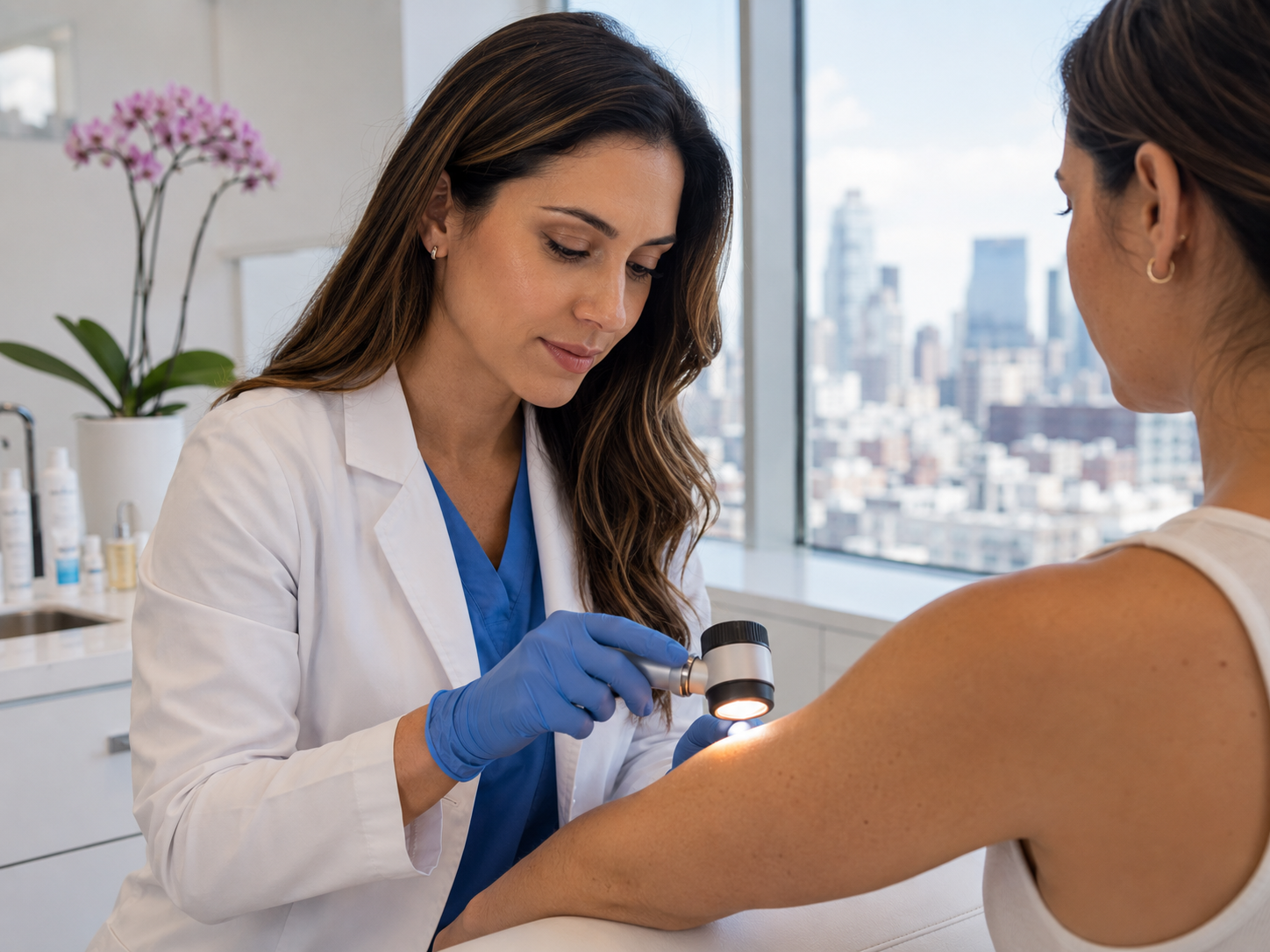
This guide ranks seven specific warning signs by urgency, from 'see a consultant within a week' to 'monitor and consider booking'. Each is grounded in the same evidence base that drives the ABCDE framework and the NICE two-week-wait criteria. Use it as a self-check tool, not a diagnostic one; only a GMC-registered consultant with dermatoscopy can confirm what a mole actually is.
Why symptom-led self-checking actually works
Pure visual self-examination has a known limitation: most patients struggle to apply the ABCDE framework objectively to their own skin. The asymmetry that strikes a dermatologist as significant often blends into a patient's general impression of 'a mole'. What patients do notice reliably is change and symptoms, the dynamic features that interrupt their normal self-image of their own skin.[1]
Studies of patients who self-detected melanoma consistently identify the same triggers: 'it grew', 'it itched', 'it bled', 'it became darker'. These aren't noticed by applying a checklist; they're noticed because something interrupted normal life. That's why the framework below is built around symptoms and changes rather than visual features held statically against a chart.
The ranking that follows is based on positive predictive value (the proportion of patients with that sign who turn out to have a clinically meaningful diagnosis). Higher up the list = higher probability of finding something on examination = more urgent to get a consultant's eye on it. Anything in the top three warrants booking within a week; anything in the bottom four warrants booking within a month or so unless multiple signs cluster together, in which case move it up.
Sign 1 (highest urgency): rapid change in size, colour or shape
Any mole that has visibly changed in the last 1-3 months should be seen by a consultant within a week. The change can be growth (the most common), darkening, the development of multiple colours, the appearance of an irregular border where there wasn't one, or the lifting of a previously flat mole. The key word is rapid: a mole that's slowly drifted across decades is normal evolution; a mole that looks different from the photo on your phone three months ago is not.
Rapid change is the strongest single predictor in melanoma self-detection studies, accounting for around 70% of self-detected cases.[1] It is also the simplest framework to apply: you don't need to know what the mole 'should' look like, you only need to know what it did look like. Phone photographs from the past few months, even casual ones, are surprisingly useful here.
Practical action. Take a current dated photograph today. If you have an older photograph, line them up side by side. If the mole has visibly changed across that gap, book a consultation. If you don't have a baseline photo, the change you've already noticed mentally is enough; trust the perception and book the appointment.
Sign 2 (high urgency): bleeding or crusting without trauma
A mole that bleeds spontaneously, crusts over without injury, or fails to heal after minor trauma should be seen within a week. Healthy moles do not bleed unless physically scraped or cut; persistent bleeding suggests the lesion's surface has lost integrity, which is more common in invasive melanoma and in non-melanoma skin cancers (basal cell and squamous cell carcinoma) than in benign naevi.
Distinguishing trauma-bleeding from spontaneous bleeding matters. A mole nicked while shaving usually bleeds briefly, scabs cleanly, and heals within a week without recurrence. A spontaneously bleeding mole bleeds without obvious cause, often more than once over weeks, and may crust and bleed cyclically. The latter is the pattern to act on.
Crusting without bleeding is the same warning in milder form: a thin crust forms repeatedly on the surface of a mole that wasn't crusted before. This is sometimes mistaken for a 'scab from nothing' and rationalised as 'I must have caught it'. Once is plausible; twice is reason to book a consultation.
Sign 3 (high urgency): persistent itching, tingling or pain
A mole that itches persistently, that develops tingling or pain, or that simply 'feels different' from the rest of the skin warrants a consultant's eye. Healthy moles do not have nerve endings that produce sensation; the perception of sensation usually means inflammation in the surrounding tissue, which can occur in atypical naevi and in early melanoma as the immune system reacts to abnormal cells.[2]
The pattern that matters is persistent, over weeks rather than minutes. A brief itch on a mole after a hot shower or insect contact is meaningless. An itch that recurs every day for two weeks, or a constant low-grade tingle, is not. Some patients describe it as 'I keep noticing this mole', a sense of awareness rather than obvious symptom; that's worth taking seriously too.
Pain is rarer but more concerning. A genuinely painful mole is uncommon and warrants urgent assessment, particularly if combined with any other sign on this list. Don't wait to see if it settles; book the consultation.
Sign 4 (moderate urgency): a new mole appearing after age 30
Most adult moles appear during childhood and adolescence and stabilise by the late twenties. After age 30, new mole formation slows considerably; after age 40 it should be rare. A genuinely new mole appearing in adulthood, particularly after age 40, deserves a consultant's eye even if it looks unremarkable on its own.
The distinction matters because melanoma sometimes presents as a new pigmented lesion rather than as a change in an existing mole. The lesion looks unremarkable to the patient (it's just 'a mole'), but it didn't exist a year ago, which is the key data point. We cover this in detail in our 'New moles after 30' guide.
Practical test. If you can't remember a specific mole from family photographs, beach holidays or routine washing in the last few years, treat it as new. Adults often have a strong sense of 'I'd have noticed that before' even without explicit memory; trust the instinct and book.
Sign 5 (moderate urgency): a mole that looks different from your others
Most people's benign moles share a family resemblance: similar size, similar shade, similar shape. The 'ugly duckling sign' is the observation that the mole that doesn't fit this pattern, the one that looks subtly different from its neighbours, is more often the one that turns out to be melanoma. Multiple validation studies have confirmed the ugly-duckling approach as a clinically useful screening method.[3]
This sign is particularly useful for patients with many moles. Working through the ABCDE framework on each one is impractical when you have 30 or 40 lesions; scanning the whole field for the lesion that looks different is fast, intuitive and reliable. The 'different' criterion can be size, colour, shape, raised-versus-flat, or just an indefinable sense of 'this one stands out'.
Practical application. Stand in front of a mirror in good light and look at the moles on a single body region (back, chest, arm). Identify the lesion that doesn't quite fit. If you can identify one, book a consultation; that's the lesion you most need a dermatoscopist to look at.
Sign 6 (moderate urgency): multiple colours within a single mole
Benign moles are usually a single shade of brown. Multiple colours within one lesion, particularly black mixed with brown, or red, blue, white or grey patches, are one of the strongest single ABCDE features for melanoma. Black or blue-grey pigment in particular suggests deeper melanocytic activity, which can correspond to deeper invasion of the skin.
White or hypopigmented patches inside an otherwise pigmented mole deserve specific mention. They can indicate regression, the body's immune system attacking part of the lesion. Regression sometimes appears in benign moles, but it can also be a marker of melanoma that has elicited an immune response, and it warrants a closer look either way.
This sign is often noticed by partners or family members rather than the patient themselves; getting another set of eyes on the moles you can't easily see (back, scalp, behind ears) increases the chance of catching multi-coloured lesions. Annual partner-led 'skin scan', particularly for patients with multiple moles, is a low-effort intervention that catches a meaningful subset of problem lesions.
Sign 7 (moderate urgency): a mole larger than 6mm with irregular features
The classical ABCDE diameter rule is that any mole larger than 6mm in diameter, roughly the size of a pencil eraser, warrants a closer look. The 6mm threshold is not a hard cut-off but a reasonable trigger: most early melanomas, by the time they become clinically obvious, have crossed it.
On its own, a mole over 6mm is a soft warning. Plenty of benign compound naevi sit at 7-8mm and have done so for decades. What converts the size from interesting to clinically meaningful is irregularity combined with size: a 6mm mole with an irregular border, asymmetric shape, multi-toned colour, or recent growth is much more concerning than a 6mm mole that's been the same uniform brown ellipse for thirty years.
Practical use. Diameter is best used as a filter rather than a diagnosis. Anything above 6mm is worth a consultant's eye; anything below 6mm that's growing is worth one too. The combination of size plus another sign on this list moves the urgency tier higher.
What to do, in order
If any one sign in the top three (rapid change, bleeding/crusting, persistent itching/pain) applies, book a consultation within a week. We see suspect-mole patients within five working days, perform consultant dermatoscopy in the same visit, and where indicated arrange same-day excision with histology. The £250 fee covers the assessment in full; histology if needed adds £325-475 plus the lab fee.
If two or more signs in the bottom four apply (new mole in adulthood, ugly-duckling, multi-coloured, large with irregularity), treat it as the same urgency: book within a week. The combination is meaningfully more concerning than any single sign alone.
If only one sign in the bottom four applies and there's no rapid change or symptom, book within a month. Continue self-examination in the meantime, and bring the appointment forward if anything changes. A delay of a few weeks for a mildly atypical-looking mole is usually fine; a delay of months is harder to defend.
Common questions
Frequently asked
References
Sources cited
- Brady MS, Oliveria SA, Christos PJ, et al. Patterns of detection in patients with cutaneous melanoma. Cancer. 2000;89(2):342-347. View source
- Bauer J, Garbe C. Acquired melanocytic nevi as risk factor for melanoma development. Pigment Cell Res. 2003;16(3):297-306. View source
- Grob JJ, Bonerandi JJ. The 'ugly duckling' sign: identification of common characteristics of nevi. Arch Dermatol. 1998;134(1):103-104. View source
- NICE guideline NG12. Suspected cancer: recognition and referral. Updated 2023. View source
- Friedman RJ, Rigel DS, Kopf AW. Early detection of malignant melanoma: the role of physician examination and self-examination. CA Cancer J Clin. 1985;35(3):130-151. View source