
Most adults carry between 10 and 40 moles. To the untrained eye they look broadly similar, small brown marks scattered across the body. To a dermatologist they are remarkably different, and the difference comes down to one question: where in your skin do the pigment-producing cells (melanocytes) sit?
That depth and arrangement is what dermatology jargon captures. A 'junctional' mole, a 'compound' mole and an 'intradermal' mole are not three random labels, they are three precise descriptions of melanocyte location. Each behaves differently over time, carries a different risk profile, and warrants a different clinical response.
This guide walks through every category a UK consultant might use about your moles, in plain English. By the end you will know what your dermatologist means when they call a mole 'compound' or 'dysplastic', and what (if anything) that classification implies for you.
A quick refresher on what a mole actually is
A mole, technically a 'melanocytic naevus', is a cluster of pigment-producing cells called melanocytes. Melanocytes are the cells that give skin its colour, manufacturing the pigment melanin and distributing it to surrounding cells. Most are scattered evenly through the skin. A mole is what happens when, for genetic and developmental reasons, a group of them clusters together in one spot.
Some moles are present from birth (congenital naevi). Most appear in childhood or early adulthood, often triggered by sun exposure or hormonal change, and stabilise by middle age. After 40, new moles become less common and any new mole appearing in older adulthood deserves a closer look.
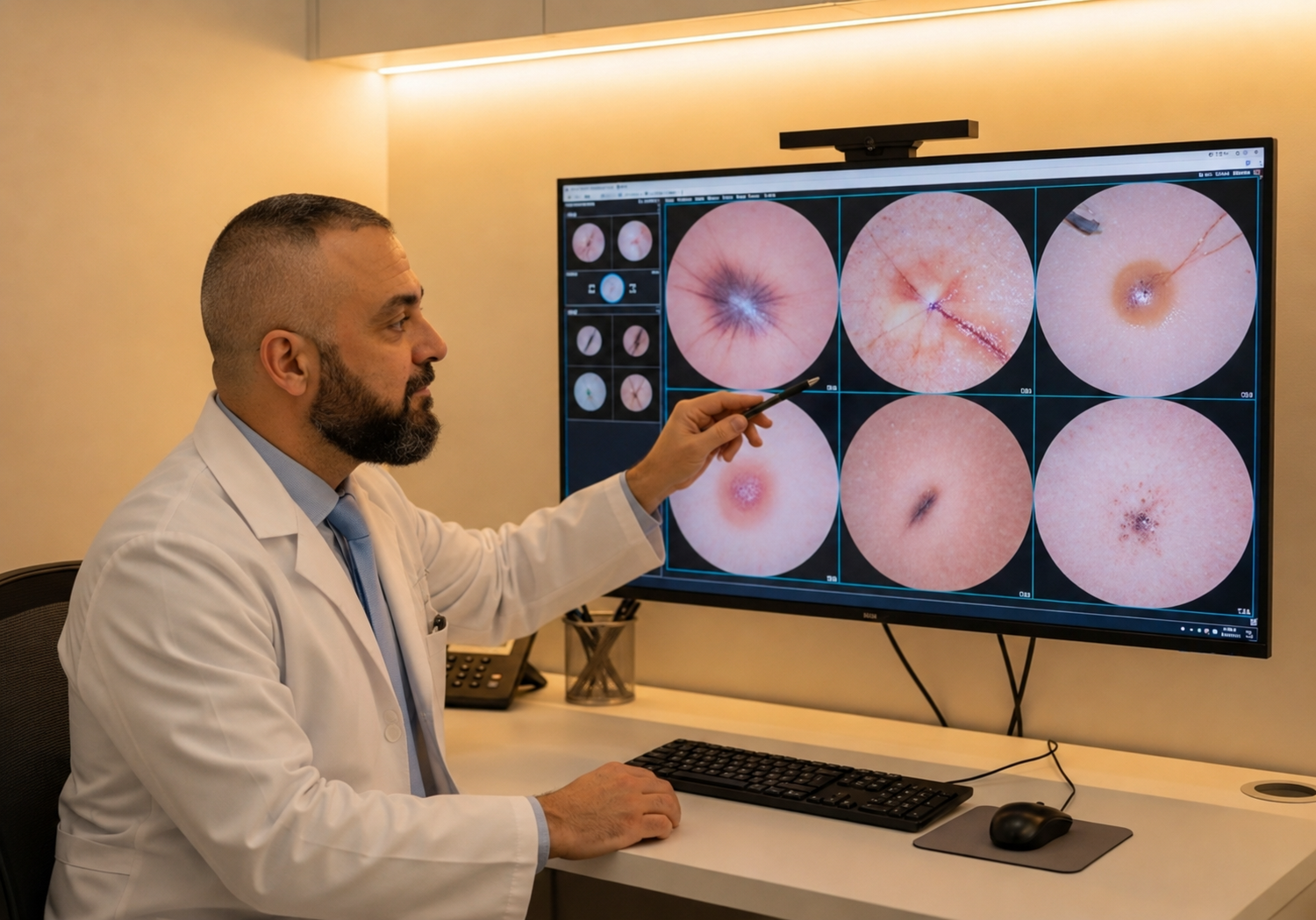
What dermatologists pay attention to is not the existence of a mole but the architecture of the cells inside it: where they sit, how they're organised, whether the arrangement is orderly or disordered. That architecture is what every other section of this guide describes.
The skin layer roadmap
To make sense of mole types, you need a working mental model of skin's layers. From the surface inward there are three: the epidermis (the top, thinner layer that forms the visible surface), the dermis (the thicker layer beneath, containing nerves, vessels and hair follicles), and the subcutis below that.
Melanocytes naturally live at the boundary between the epidermis and dermis, the so-called 'dermo-epidermal junction'. When a mole forms, the clustered melanocytes can stay at that junction, drift up into the epidermis, drop down into the dermis, or spread across both. Where they end up determines the mole's clinical name and its likely behaviour.
All of the categories below are descriptions of one thing, the depth distribution of the melanocyte cluster. None of them inherently makes a mole 'good' or 'bad', but each carries a typical risk profile and a typical visual appearance.
Junctional naevi
A junctional naevus is the simplest configuration: melanocytes clustered exactly at the dermo-epidermal junction, neither rising into the epidermis nor descending into the dermis. They are the typical mole of childhood and adolescence, although adults retain plenty of them.
Visually they tend to be flat (because the cluster does not bulge upward), evenly brown to dark brown in colour, and well-defined at the edge. Most are small, under 5mm, and round or oval. Run your fingertip across one and you should not feel a noticeable bump.
Junctional naevi are almost always entirely benign. They are the kind of mole most patients have without realising, and they require no special action unless something about them is changing. The ABCDE framework still applies: any junctional mole that becomes asymmetric, develops irregular borders, varies in colour, grows past 6mm or changes over time deserves a consultant's eye.
Compound naevi
Compound naevi are the most common adult mole. Here, the melanocyte cluster spans both sides of the dermo-epidermal junction: some cells sit at the junction itself, others have descended into the upper dermis. The dual location explains the typical clinical appearance.
Compound naevi are usually slightly raised, just enough to feel under a fingertip. They tend to be brown, with relatively soft texture, and the border is typically clean. Many will have a faint surface texture (a slight cobblestone feel) because the cluster's depth varies. Diameter is usually under 6mm but can be larger.
Like junctional naevi, the overwhelming majority of compound naevi are benign. They are the bread and butter of mole assessment, and most patients hearing the word 'compound' from their consultant should hear it as reassurance: it is the most common, expected configuration. Watching for ABCDE change still applies, particularly as compound moles can in rare cases evolve over decades.
Intradermal naevi
An intradermal naevus has the melanocyte cluster wholly within the dermis, with no remaining presence at the dermo-epidermal junction. In practice this is the typical mole of middle age and beyond: a junctional mole that drifted, then a compound mole that finished its descent, leaving the cluster entirely in the deeper layer.
Visually, intradermal naevi are often quite different from their junctional and compound predecessors. They tend to be raised and dome-shaped, sometimes pedunculated (sitting on a thin stalk), and are commonly pale pink, skin-coloured, or only faintly brown. The pigment is masked because the melanocytes sit deeper in the skin and the overlying tissue softens the colour.
Intradermal naevi are very low-risk lesions. Their melanocytes have effectively 'matured' and the architecture is stable. The reason patients sometimes ask about them is cosmetic, they may catch on clothing, sit in a visible facial position, or be irritated by shaving. Cosmetic shave excision is straightforward in those cases.
Dysplastic and atypical naevi
'Dysplastic' is where the language shifts from 'where the cells sit' to 'how the cells are organised'. A dysplastic naevus, sometimes called an atypical naevus clinically, is a mole whose internal architecture is disordered: melanocytes are unevenly distributed, the boundary between mole and surrounding skin is less clear, and individual cells may show subtle abnormalities visible only under a microscope.
Dysplastic naevi are usually compound in their depth distribution but their visual signature is different. They tend to be larger than typical (>6mm), have an irregular border, and show varied colour, the same features the ABCDE framework flags. They are not melanoma, but they sit on a continuum that includes melanoma at one end.
What matters clinically is what dysplastic naevi tell us about baseline risk. People with dysplastic naevi have a meaningfully higher lifetime risk of melanoma, both within the dysplastic moles themselves and elsewhere on the skin.[1] The right response is not necessarily to remove every one, that would be impractical for most patients, but to monitor them carefully, ideally with mole mapping, and to lower the threshold for excising any that change.
Special cases worth knowing about
Beyond the four main categories above, dermatologists recognise several specific subtypes that come up often enough to be worth a brief overview.
Congenital naevi are present from birth or appear in the first months of life. Most are small (<1.5cm) and behave like ordinary moles. Larger congenital naevi (>20cm in adulthood, classed 'giant'), are uncommon but carry a higher lifetime melanoma risk and warrant specialist surveillance from childhood.[2]
Blue naevi are small, dome-shaped, and surprisingly blue or blue-grey because their melanocytes sit deep in the dermis (the depth shifts the perceived colour). Most are entirely benign, but the unusual colour means dermatoscopy is wise to confirm.
Spitz naevi typically appear in children and adolescents as fast-growing, pink or reddish dome-shaped lesions. Histologically they share features with melanoma, and even experienced dermatopathologists sometimes find them difficult to call. Most are benign but excision and histology are commonly recommended.[3]
Halo naevi are ordinary moles surrounded by a ring of depigmented skin, the body's immune system effectively attacking the mole. Most are entirely benign and resolve over months to years. Multiple halo naevi or rapid changes warrant a consultant's eye.
What this classification actually means for you
For most moles on most patients, the type is interesting context but does not change anything you need to do. Junctional, compound and intradermal naevi are all standard, expected configurations of benign melanocytes. Knowing the label is reassuring, not actionable.
Where classification matters is when 'dysplastic' enters the conversation, when a consultant recommends ongoing monitoring or excision, or when the appearance of a mole does not match the type your dermatologist would expect for your age and skin. A mole that looks intradermal in someone in their twenties, for example, is unusual; a mole that looks junctional in someone in their seventies and is new, is a flag.
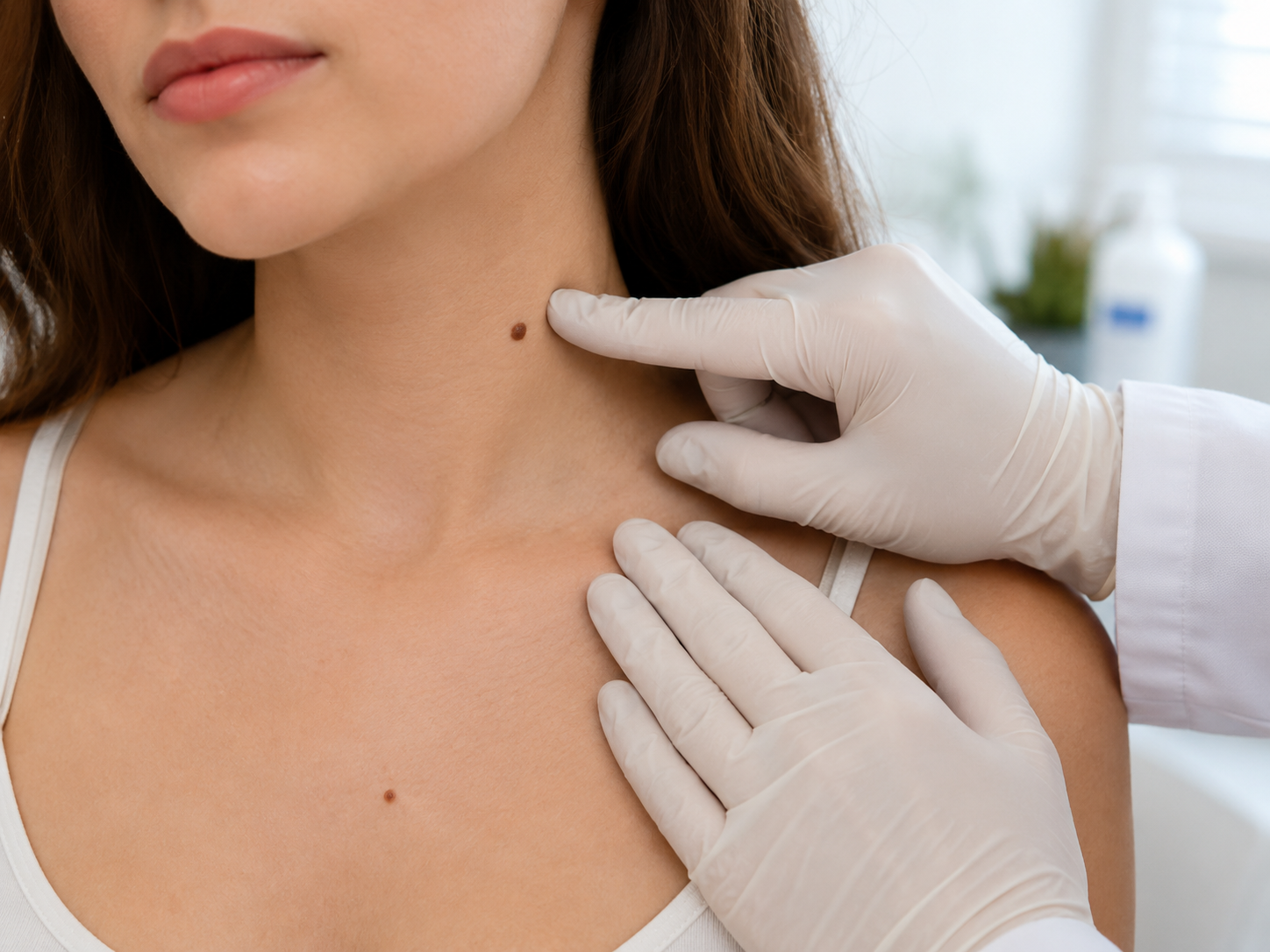
The right response in any of these cases is not panic. It is a 30-minute mole check with a GMC-registered consultant who can examine the lesion with dermatoscopy, place it accurately in this taxonomy, and tell you in plain English what (if anything) needs to happen next. Most often the answer is 'nothing'. When it isn't, you'll leave with a clear plan rather than a nagging worry.
Common questions
Frequently asked
References
Sources cited
- Naeyaert JM, Brochez L. Clinical practice. Dysplastic nevi. N Engl J Med. 2003;349(23):2233-2240. View source
- Krengel S, Hauschild A, Schäfer T. Melanoma risk in congenital melanocytic naevi: a systematic review. Br J Dermatol. 2006;155(1):1-8. View source
- Luo S, Sepehr A, Tsao H. Spitz nevi and other Spitzoid lesions: part I. Background and diagnoses. J Am Acad Dermatol. 2011;65(6):1073-1084. View source
- Bauer J, Garbe C. Acquired melanocytic nevi as risk factor for melanoma development. A comprehensive review of epidemiological data. Pigment Cell Res. 2003;16(3):297-306. View source