

Receiving a histology report is a strange experience. It's a 1-2 page document that arrives 7 to 10 days after your excision, usually by post and email, full of language that reads like a foreign clinical dialect: 'compound naevus with focal architectural disorder', 'Breslow thickness 0.4mm', 'margins clear', 'mitotic rate 1/mm²'.
Each of those terms has a specific meaning that determines exactly what (if anything) needs to happen next. A patient who can read their own report walks into the follow-up consultation already understanding the answer; the appointment becomes confirmation rather than translation.
This guide translates the four fields a UK histology report contains, plus the additional features that show up on more complex reports. By the end you'll know what 'Breslow thickness' is, what 'clear margins' means, and what to do when the report mentions 'positive' or 'involved' margins. Written and medically reviewed by a GMC-registered consultant who specialises in melanoma surgery.
What histology actually is
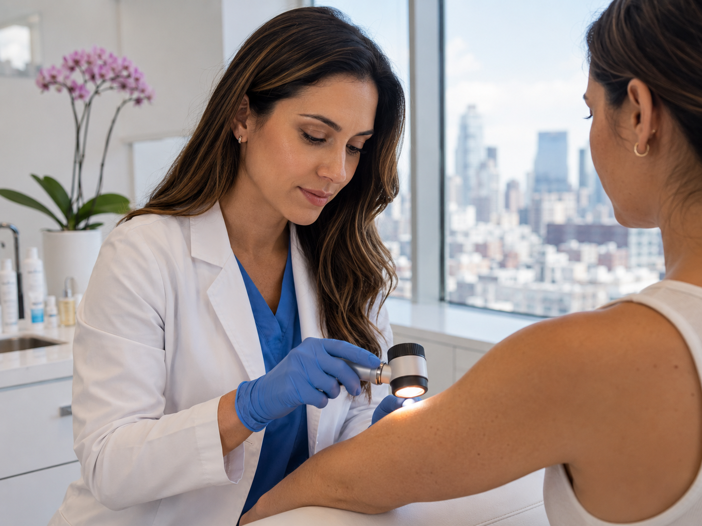
Histology is the microscopic examination of tissue. After your lesion is removed, the specimen is placed in a fixative (typically formalin), transported to a UKAS-accredited pathology lab, and processed: dehydrated, embedded in paraffin wax, sliced into thin sections, mounted on glass slides, and stained (most commonly with haematoxylin and eosin, the pink-and-purple staining most people picture when they think of pathology).
A consultant histopathologist then examines the slides under a high-magnification microscope, comparing the cellular architecture against thousands of reference patterns from training and practice. The pathologist describes what they see, makes a diagnosis, measures relevant features, and signs off the report. The whole process typically takes 5 to 10 working days from when the lab receives the specimen.
What you receive at the end is the pathologist's signed report. It looks technical because it is, but the structure is consistent: diagnosis, cell type, depth, margins, and any other features that influence staging or follow-up.
The diagnosis field, what the lesion actually is
The first and most important field is the diagnosis itself. UK histology reports use one of three categories.
Benign: the lesion is a common naevus, intradermal, junctional, compound, blue, Spitz, halo, or one of the less common benign types. The cells are not malignant. No further action is needed beyond healing and ongoing self-monitoring of your remaining moles. The vast majority of cosmetically excised moles fall into this category.
Atypical or dysplastic: the architecture is disordered relative to a routine benign naevus, but the cells are not malignant. The pathologist will usually grade severity (mild, moderate, severe). Mild and moderate atypia with clear margins generally need no further action. Severe atypia close to or touching the margin may warrant a wider local excision.
Malignant: the lesion contains cancer cells. The most common malignancies seen on routine excision are basal cell carcinoma (BCC), squamous cell carcinoma (SCC), and melanoma. Each has its own management pathway, see the cell type section below for what each means clinically.
The cell type field, which kind of cells
If the diagnosis is malignant, the next field tells you the cell type. This determines the next step entirely.
Basal cell carcinoma (BCC) is the most common skin cancer and usually the most treatable. It tends to grow locally rather than spread, and complete excision with clear margins is curative in the great majority of cases. BCCs are subtyped (nodular, superficial, infiltrative, micronodular, morphoeic) and the subtype affects how aggressive the surgical approach needs to be.
Squamous cell carcinoma (SCC) is less common but more behaviourally aggressive than BCC. Most early SCCs are still cured by complete excision, but a minority can spread, particularly large or deeply invasive lesions on the lip, ear, or pre-existing scar. Histology depth and differentiation grade guide whether further imaging or sentinel-node assessment is needed.[1]
Melanoma is the cancer dermatology pays most attention to. The pathologist will report subtype (superficial spreading, nodular, lentigo maligna, acral lentiginous), depth (Breslow thickness, see next section), ulceration, mitotic rate, and several other features that together determine staging and treatment. A melanoma diagnosis triggers a multidisciplinary team discussion before next steps are agreed.
The depth field, Breslow thickness explained
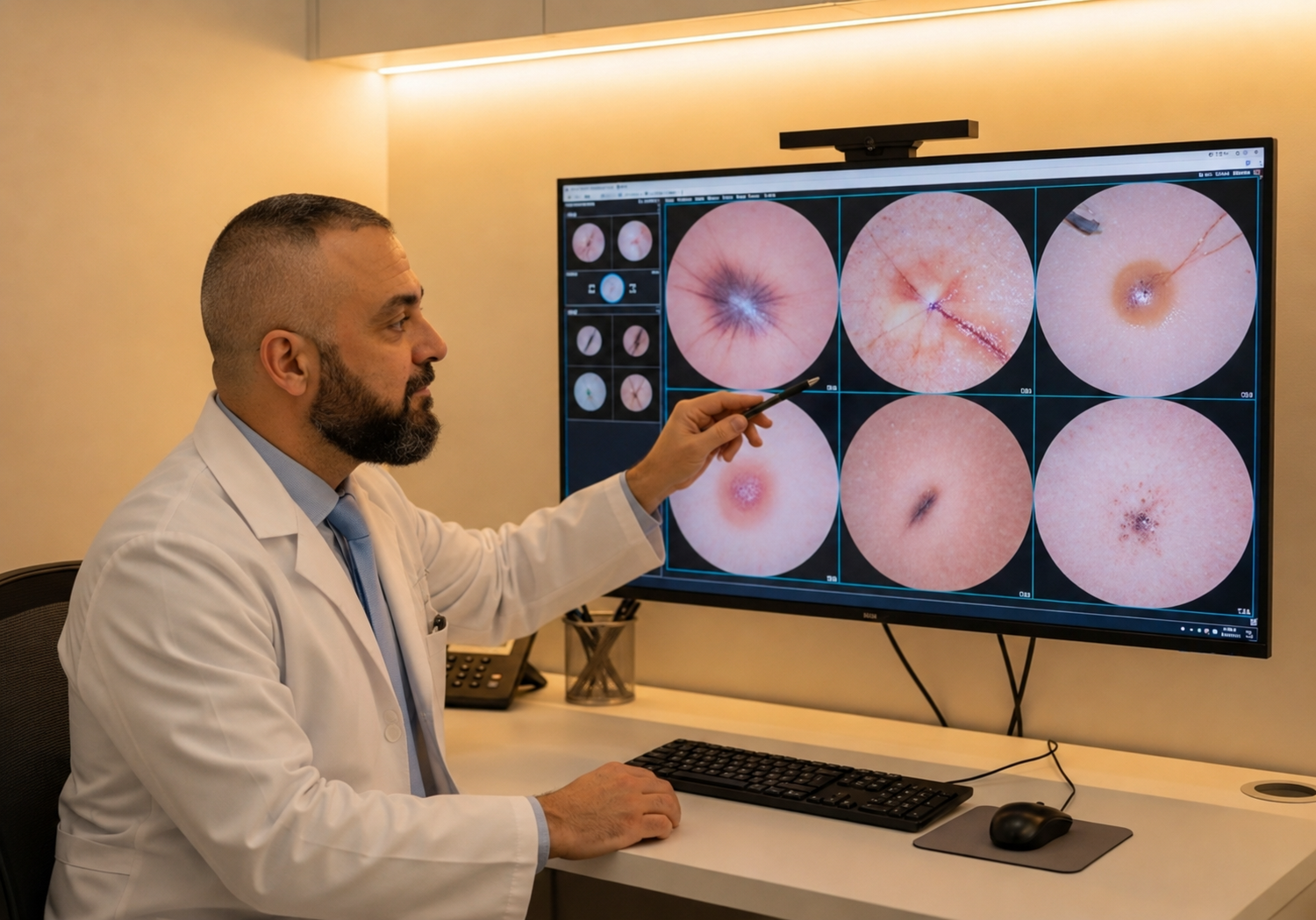
For melanoma specifically, depth is the strongest single predictor of outcome. The pathologist measures the depth of the tumour in millimetres, from the top of the skin to the deepest tumour cell, using a calibrated optical micrometer. This measurement is called the Breslow thickness, named after Alexander Breslow who introduced it in 1970.[2]
The thresholds that drive management are well-defined: ≤0.8mm (very thin, often curative with appropriate margins), 0.8-2mm (intermediate, sentinel lymph node biopsy commonly recommended), 2-4mm (deeper, sentinel-node biopsy and possibly imaging), >4mm (deep, more aggressive workup). The exact thresholds shift slightly with each AJCC staging update; what doesn't shift is that thinner is better.
Some reports also mention Clark level, an older anatomical measure (I = epidermis only, II-V increasingly deep). Clark level has been largely superseded by Breslow thickness in modern UK practice but you may still see it on reports, particularly for thinner melanomas where it provides additional context. For BCC and SCC, Breslow doesn't apply; depth is reported anatomically (e.g. 'invasion to mid-dermis').
The margins field, was it all removed
Margins describe whether the lesion was fully removed with a clean border of normal skin around it. The pathologist examines the edges of the specimen and reports one of three results.
Clear margins: the tumour does not reach the edge of the specimen, with a measurable distance between the deepest tumour cell and the closest cut edge. The report will usually quote the closest distance (e.g. '1.5mm'). This is what you want.
Close margins: the tumour reaches close to but not at the cut edge. Whether this matters depends on the tumour type. For BCC, close margins (<1mm) often warrant wider re-excision. For melanoma, the surgical guideline already specifies the margin to take based on Breslow thickness, so 'close' typically means the planned margin was achieved exactly.
Involved or positive margins: the tumour reaches the cut edge of the specimen, meaning some tumour was likely left behind. A wider local excision is typically recommended to remove the residual tumour and re-establish clear margins. This is a straightforward outpatient procedure under local anaesthetic, not a major operation.
Other features: mitoses, ulceration, regression
Beyond the four core fields, more complex reports include several additional descriptors that can shift staging and follow-up. They are most relevant for melanoma but appear on other reports too.
Mitotic rate counts how many cells are visibly dividing per square millimetre of tumour. Higher mitotic rates indicate faster proliferation and often more aggressive behaviour. For thin melanomas (<1mm) the mitotic rate is one of the factors that influences whether sentinel-node biopsy is offered.
Ulceration describes whether the surface of the tumour has broken through the overlying epidermis. Ulcerated melanomas behave more aggressively than non-ulcerated ones at the same Breslow depth, so the staging system explicitly bumps up.
Regression describes histological evidence that the immune system has attacked part of the tumour, leaving behind fibrotic tissue and inflammatory cells where tumour used to be. Regression sometimes appears in benign moles, but in melanoma it can indicate the tumour was thicker than the current Breslow measurement suggests, because some of the tumour has been destroyed.
Perineural or lymphovascular invasion describes whether the tumour has invaded into nerve sheaths or blood/lymph vessels respectively. Both are markers of more aggressive behaviour and influence the surgical and surveillance plan.
What happens after a malignant result
If your histology shows BCC, SCC or melanoma, your consultant will call you with the result, usually the same day the report is received, and arrange a follow-up appointment to walk through the implications and agree the next step.
For most early BCCs with clear margins, no further action is needed beyond ongoing skin surveillance. For SCC, the treating consultant will assess whether further imaging or excision is warranted based on size, depth and location. For melanoma, the case is brought to a multidisciplinary team meeting (MDT) involving dermatology, plastic surgery, and where appropriate oncology, to agree the surgical plan (typically a wider local excision based on Breslow depth) and whether sentinel-node biopsy is appropriate.[3]
Where appropriate we coordinate referral into NHS specialist services so you're not navigating the system alone. The transition is structured and warm, not a discharge with a leaflet. Ongoing surveillance, annual mole checks or full mole mapping, is recommended after any skin cancer diagnosis because the risk of a second primary lesion is meaningfully elevated for the rest of life.
Common questions
Frequently asked
References
Sources cited
- Stratigos AJ, Garbe C, Dessinioti C, et al. European interdisciplinary guideline on invasive squamous cell carcinoma of the skin: Part 1. epidemiology, diagnostics and prevention. Eur J Cancer. 2020;128:60-82. View source
- Breslow A. Thickness, cross-sectional areas and depth of invasion in the prognosis of cutaneous melanoma. Ann Surg. 1970;172(5):902-908. View source
- Marsden JR, Newton-Bishop JA, Burrows L, et al. Revised UK guidelines for the management of cutaneous melanoma 2010. Br J Dermatol. 2010;163(2):238-256. View source
- Royal College of Pathologists. Standards and datasets for reporting cancers: Dataset for histopathological reporting of primary cutaneous malignant melanoma and regional lymph nodes. 2019. View source
- Gershenwald JE, Scolyer RA, Hess KR, et al. Melanoma staging: Evidence-based changes in the American Joint Committee on Cancer eighth edition cancer staging manual. CA Cancer J Clin. 2017;67(6):472-492. View source