

Many UK clinics offer laser mole removal. It is marketed as quick, scarless, easy, all the right adjectives for a cosmetic decision. Patients ask about it constantly, often surprised when we explain we do not offer it.
We do not use laser for moles. The reason is clinical, not stylistic: laser destroys the tissue that pathology depends on, and even moles that look entirely benign on the surface can hide diagnostic surprises underneath. Once a mole is lasered, the option to send it for histology is gone forever.
This guide explains exactly why, what the alternatives are, and why a deliberate, slightly less convenient technique choice is the safer one across every clinical scenario, including purely cosmetic cases. Written and medically reviewed by a GMC-registered consultant plastic surgeon who performs hundreds of mole removals each year.
What laser actually does
Laser mole removal works by aiming a focused beam of light at the mole. The pigment in the mole (melanin) absorbs the laser energy preferentially, heating up rapidly and breaking down. With repeated passes, the visible mole fades. From the patient's perspective the result looks tidy, the surface skin remains intact, and the recovery is fast.
The clinical problem is what happens at the cellular level. The laser energy vaporises the pigmented cells, which is to say it destroys them. Nothing is preserved. There is no specimen left to send to a pathology lab. The melanocytes that made the mole, the architecture they sat in, the boundary between mole and normal skin, all of it is gone.
For an entirely benign cosmetic mole, you might reasonably argue this doesn't matter. The mole was harmless, you wanted it gone, the result is good. The issue is that you cannot reliably know in advance which moles are entirely benign. Naked-eye examination is good but not perfect. Dermatoscopy improves accuracy but doesn't eliminate uncertainty. The only definitive answer is histology, and histology requires a specimen.
Why histology matters even for cosmetic cases
Around 1 to 2% of moles that look benign on dermatoscopic examination turn out to have unexpected histological findings.[1] Most of those findings are mild atypia that don't change the patient's outlook. A small subset, perhaps 1 in 1,000 cosmetic excisions, are found to harbour an unexpected melanoma in situ or even early invasive melanoma.
When that 1-in-1,000 finding occurs, the value of having sent the specimen to histology is enormous. It triggers wider excision, MDT review, and surveillance, all of which materially improve the patient's outcome. When the same mole is lasered instead, the diagnosis is missed; the patient leaves believing they have removed a benign mole, when in fact they have destroyed evidence of an early melanoma. The melanoma cells that survived deeper in the skin are then free to progress, undetected, until they re-present years later as a much more dangerous lesion.
This is the case against laser in a single sentence: it works perfectly for the 999-in-1,000 patients who don't need histology, and catastrophically fails the 1-in-1,000 patient who did. We don't know in advance which patient is which. The only safe approach is to treat every mole removal as one that might need histology, and use techniques that preserve the option.
The alternatives we use
There are four mole-removal techniques we routinely use, each chosen for the lesion it suits best. None of them sacrifices the option of histology.
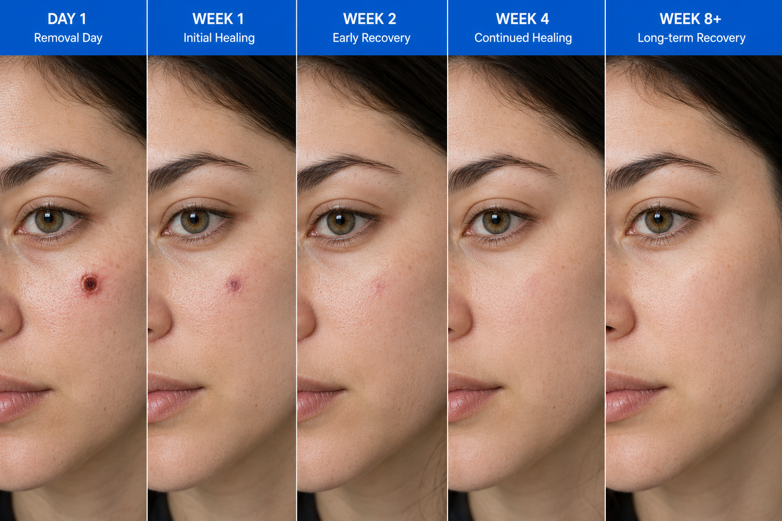
Shave excision trims the raised portion of a mole flush with the surrounding skin using a fine surgical blade. The trimmed tissue is preserved as a specimen and can be sent to histology. Shave excision suits raised, dome-shaped benign moles where a vertical sample of the upper part of the lesion is sufficient. Healing is fast, no stitches are required, and the resulting flat pale circle fades over weeks.
Full-thickness elliptical excision removes the entire lesion plus a small margin of normal skin around it, in one clean ellipse, sutured closed with fine sutures. The complete specimen, including margins, goes to histology. This is the technique of choice for any lesion needing definitive pathology, anything dermatoscopically suspicious, anything a patient wants thoroughly checked, anything flat or deep where a shave wouldn't capture the full lesion.
Electrocautery uses precise heat to remove very small superficial lesions. Skin tags, cherry angiomas, and small fibroepithelial polyps respond well. The specimen is altered by the heat but a limited histological examination remains possible, and the technique suits lesions where the pre-test probability of an unexpected finding is genuinely low.
Cryotherapy uses a brief application of liquid nitrogen to freeze and destroy superficial lesions. We use it sparingly because, like laser, it does not preserve a specimen. We reserve it for narrowly defined cases where the lesion is unambiguously benign and superficial.
The scar trade-off
The most common patient question after we explain the no-laser policy is: but won't I have a worse scar with surgical excision? It is a fair question, and the answer is more nuanced than 'yes' or 'no'.
A shave excision typically leaves a flat pale circle of slightly different texture, a scar that fades substantially over months to years and is often invisible from a normal conversational distance after a year. A full-thickness excision leaves a thin linear scar in the direction of the natural skin tension lines, which similarly fades over 6 to 12 months when sun-protected. Neither is invisible. Neither is dramatic.
Laser, when it works as advertised, can leave essentially no scar. When it doesn't (and it often doesn't, particularly for darker skin types where the pigment-targeting mechanism is less precise), it can leave irregular hypopigmented or hyperpigmented patches that are more visible than a clean surgical scar. Skilled surgical excision plus diligent post-removal scar care produces an outcome that is usually better than reasonable laser, and not noticeably worse than ideal laser, with the diagnostic option preserved.[2]
When laser is the right tool
We are not anti-laser as a technology. Laser is excellent for several dermatology applications: vascular lesions like port-wine stains and spider veins, photo-rejuvenation, hair removal, and treatment of certain pigmentation conditions like solar lentigines that have been definitively diagnosed.
What laser is wrong for is melanocytic naevi, the pigmented lesions formed by clusters of melanocytes that we collectively call moles. The reason is the diagnostic-evidence argument above, not any property of the laser itself. A patient who is offered laser for a definite skin tag, cherry angioma, or other clearly diagnosed non-mole lesion can reasonably accept; the histological argument doesn't apply because the diagnosis is already settled.
If you are unsure whether a clinic is offering you laser for a mole or for a different lesion type, ask explicitly: 'Is this a melanocytic naevus, and if so, are we sending it to histology?' If the answer is yes-then-no, find a different clinic.
What this means for your appointment
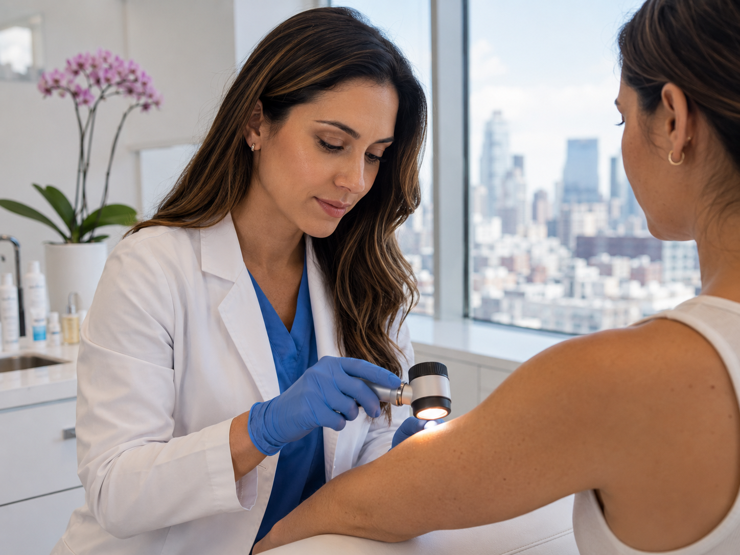
When you book a cosmetic mole removal with us, the appointment runs in three parts. First, dermatoscopic examination of the lesion to confirm it is appropriate for cosmetic-only removal. Second, a discussion of which non-laser technique gives the best outcome for that specific lesion: shave for raised benign moles, full-thickness excision for flat or deeper moles, electrocautery for small superficial benign growths. Third, the procedure itself, performed in the same visit under local anaesthetic.
If the dermatoscopic examination flags any concern, we will recommend switching to a Skin Cancer Check (£250) and routing the lesion through histology, regardless of how cosmetic the original brief was. Your safety always takes priority over the original appointment plan.
The trade-off is genuine: a slightly less convenient technique, a slightly longer recovery, and a small visible scar in exchange for the diagnostic certainty that nothing dangerous was destroyed in the process. For everyone we have treated this way, that trade-off has been worthwhile. It is the trade-off the dermatology profession has standardised on for the right reasons.
Common questions
Frequently asked
References
Sources cited
- Argenziano G, Albertini G, Castagnetti F, et al. Early diagnosis of melanoma: what is the impact of dermoscopy? Dermatol Ther. 2012;25(5):403-409. View source
- Sardana K, Chakravarty P, Goel K. Optimal management of common acquired melanocytic nevi (moles): current perspectives. Clin Cosmet Investig Dermatol. 2014;7:89-103. View source
- British Association of Dermatologists. Position statement on the use of lasers and intense pulsed light for cosmetic purposes. Br J Dermatol guideline series. View source
- Carlson JA, Slominski A, Linette GP, Mihm MC Jr, Ross JS. Biomarkers in melanoma: predisposition, screening and diagnosis. Expert Rev Mol Diagn. 2008;8(3):281-302. View source