
The single most common reason patients put off a mole check that would reassure them is not knowing what to expect. There's a vague sense the appointment will be awkward, that they will need to undress fully, that the consultant might find something dramatic and rush them off to surgery, that the whole process is somehow more medical than it actually is.
It isn't. A standard mole consultation is 30 minutes, structured, and almost always ends with reassurance. The minority of appointments that don't end that way still end with a clear, calm next step rather than a scramble. This guide walks through every stage in order so you arrive knowing exactly what's coming.
Written by a GMC-registered consultant who runs these appointments daily. Tested against the experience patients describe afterwards, the real-world appointment is consistently easier than the version people imagine before they walk in.
Before your appointment
Preparation for a mole check is light. There is no fasting, no scans to bring, no GP referral required, and no pre-appointment paperwork beyond the brief health declaration you'll fill in on arrival. Most patients book online and turn up.
Two practical things help. First, take dated phone photographs of any mole you've noticed changing, even if the change feels small. Memory is unreliable for mole appearance, but a photograph from three months ago is hard data. Second, have a mental list of any moles you specifically want examined. Listing them at the start gives the consultant a clear focus and ensures none of them get missed.
On the practical side: avoid makeup, fake tan or nail polish on the area you want examined, since each can mask the surface appearance of pigmented lesions. Wear something easy to adjust, a button-up shirt or a t-shirt for back checks, loose trousers or shorts if you have lesions on the legs. We have gowns available for full-body work; we do not photograph anywhere covered by underwear except on specific clinical request.
Arrival, the first five minutes
You'll be greeted at reception, asked to confirm your contact details, and handed a brief health declaration form. The form covers things like medications, allergies, previous skin cancer history and any medical conditions affecting clotting. It takes most patients three minutes to complete.
If you're due to pay on the day, you'll settle the £250 mole check fee at this point (we accept card, Apple Pay, Google Pay and bank transfer). The £50 deposit you put down at booking is deducted, so the remainder is £200. There is no insurance paperwork to navigate, we are a self-pay clinic and every patient pays the same published fee.
Then you'll be shown to the consulting room. The room is private, climate-controlled, with appropriate lighting for skin examination. A chaperone is available on request and you can ask for one at any point in the appointment.
The clinical history, minutes 5 to 10
Your consultant will start with a structured but conversational history. Expect questions about: any specific moles you have noticed (this is when your phone photographs are useful), family history of melanoma or other skin cancers, your sun-exposure pattern across childhood and adulthood, any episodes of severe sunburn, your skin type (Fitzpatrick I-VI), and any previous skin lesions you've had removed.
There are no wrong answers, and no judgement. Sun exposure is a major melanoma risk factor and a holiday history of frequent unprotected sun is useful information, not something to apologise for. Be honest about it; the more accurate the history, the more usefully the consultant can weight what they find on examination.
If you've been referred or self-referred specifically about one mole, this is the right moment to describe it: when you first noticed it, what has changed, whether it itches or bleeds, whether anyone else has commented on it. Take your phone out and show the photographs.
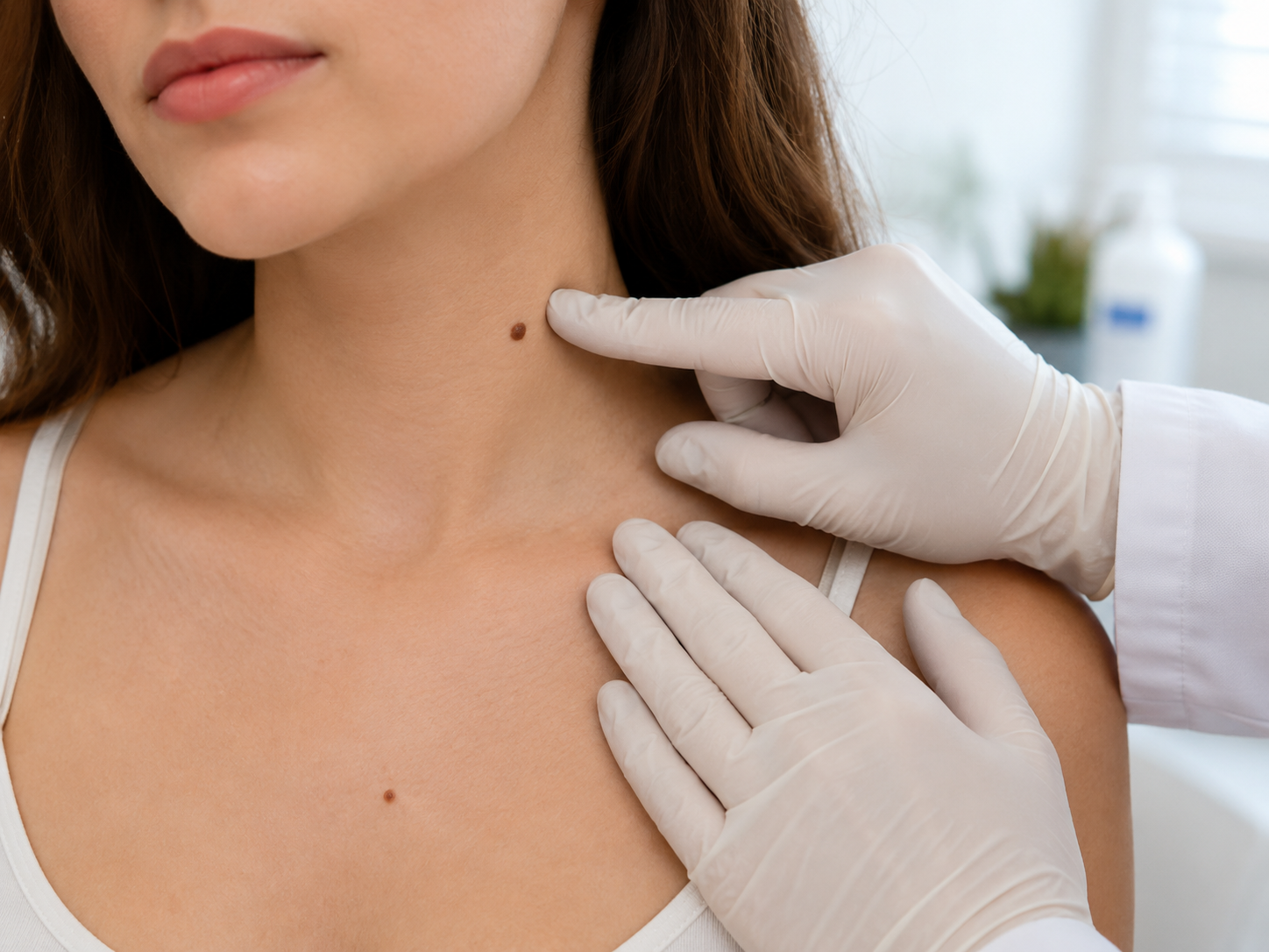
The naked-eye examination, minutes 10 to 15
The consultant will then examine the moles you have flagged, plus any others worth a closer look. Naked-eye examination is more useful than people expect: a trained dermatologist can pick up patterns of asymmetry, border irregularity and colour variation that the patient hasn't noticed. The structured surrounding-skin scan often surfaces lesions the patient didn't know existed.
You will not be asked to undress fully. The standard mole check examines the lesions you have flagged plus surrounding skin; full-body examination is offered if the consultant judges it useful or you specifically request it, and a chaperone is required for any examination of intimate areas. Underwear stays on throughout.
If the consultant flags an additional lesion you hadn't noticed, they'll explain why it caught their eye and add it to the list for dermatoscopy. This is one of the highest-value parts of a professional mole check, the moles you don't notice are exactly the ones a trained eye is most likely to catch.
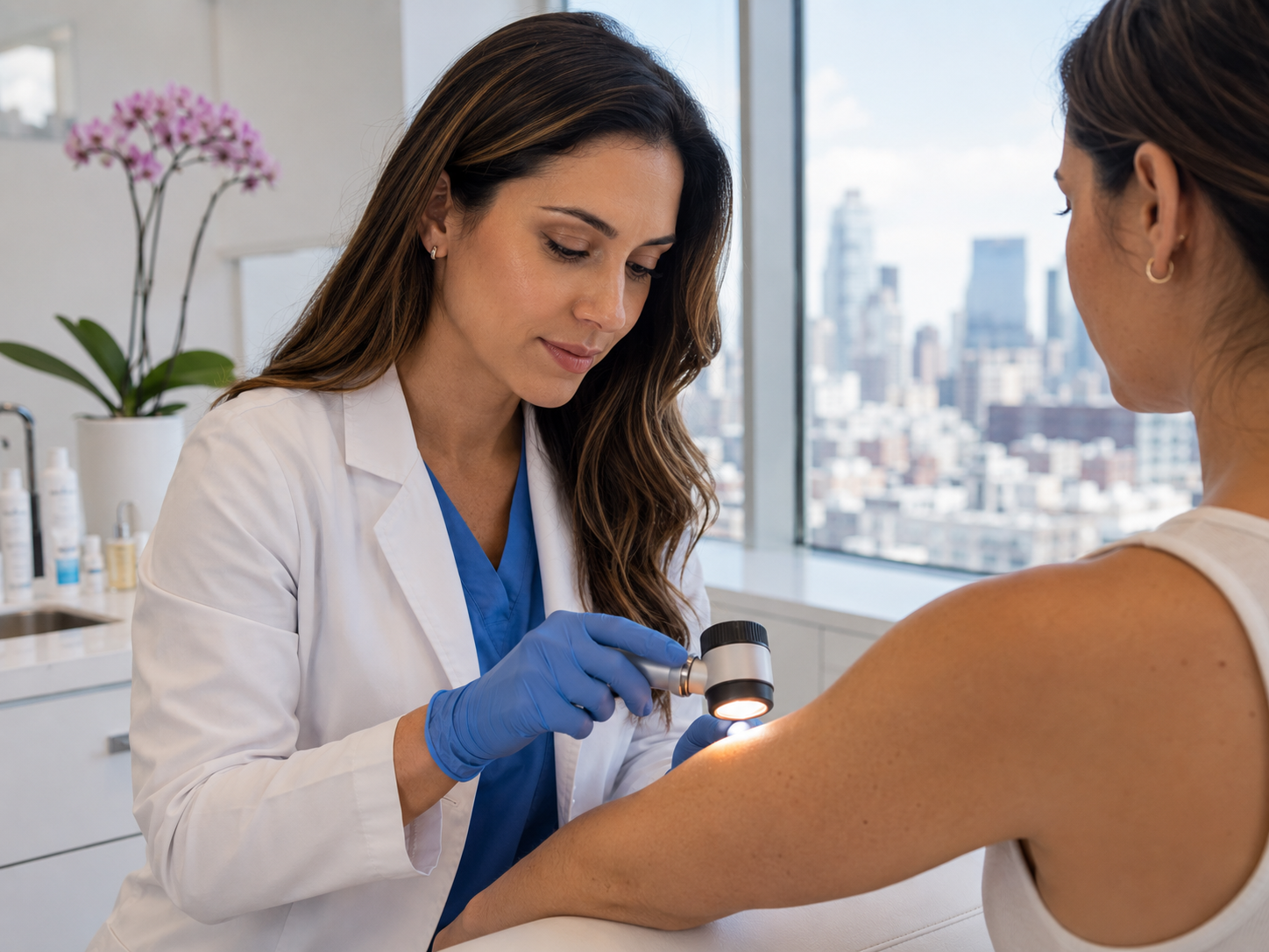
Dermatoscopy, minutes 15 to 25
Each lesion of concern is then examined with a polarised dermatoscope, a hand-held magnifier with cross-polarised light that eliminates surface reflection and reveals pigment networks, vascular patterns and structural features sitting just below the visible skin surface. This is where naked-eye examination becomes properly diagnostic.
Your consultant will hold the dermatoscope gently against the skin (it does not penetrate or hurt), examine each mole for 30 to 60 seconds, and describe what they see using the ABCDE framework. Asymmetry, Border, Colour, Diameter, Evolution. Most consultants will turn the dermatoscope's screen toward you and walk you through the features they're seeing, this is genuinely informative and worth paying attention to.
Studies repeatedly show that dermatoscopy plus expert eye outperforms expert eye alone, particularly for catching early, structurally subtle melanomas that haven't yet developed obvious clinical signs.[1] It is the diagnostic tool that separates a mole check from a glance.
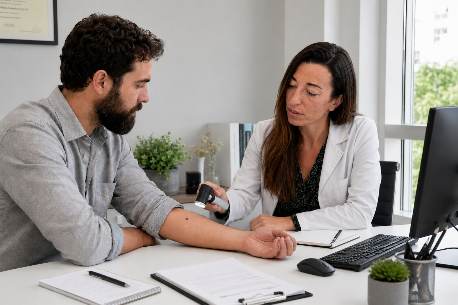
Findings and plan, minutes 25 to 30
The appointment closes with a structured discussion. Your consultant walks through what they saw on each lesion of concern, places it in plain language (benign, borderline, suspicious), and recommends a next step. The recommendation will be one of five: reassure (no action), monitor (re-photograph at 3-6 months), cosmetic removal (the lesion is benign but you'd like it gone), excise with histology (suspicious enough to warrant pathology), or refer for mole mapping (high baseline risk warranting photographic surveillance).
Whatever the finding, you should leave the appointment with a clear plan rather than a list of unanswered questions. If anything is unclear, ask. The consultant has another 30 minutes booked after yours; there's no rush, and asking questions improves the outcome.
Within 24 hours, a written report arrives by email. With your consent, it is also sent to your GP for their records, useful for continuity if a future GP wants context, and helpful if NHS dermatology referral becomes appropriate later.
Same-day removal, when it happens and what's involved
If a lesion is dermatoscopically suspicious, the consultant may recommend same-day excision. This is offered, not imposed; you can think about it and book a separate appointment if you'd prefer. Most patients accept the same-day option because it shortens the diagnostic loop.
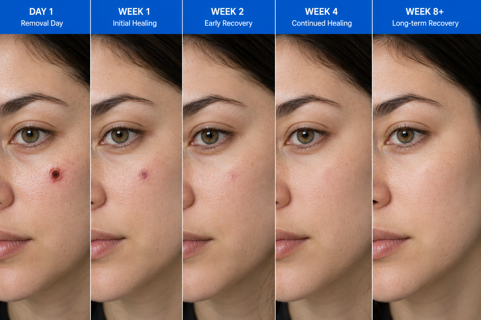
Practically: same-day excision adds 30 to 45 minutes to the appointment. The area is numbed with a small injection of local anaesthetic, the consultant performs a full-thickness elliptical excision (lesion plus a clean margin in one ellipse), and the wound is closed with fine sutures. You'll leave dressed, with written aftercare and a stitch-removal appointment booked in.
The tissue is sent same-day to a UKAS-accredited histopathology lab. Results are typically returned within 7 to 10 working days. Your consultant calls you with the result and posts a written report by recorded delivery, copied to your GP with your consent.
After your appointment
Most patients leave with reassurance and a recommendation to repeat the check at the appropriate interval (every 2-3 years for low-risk patients, annually for moderate risk, six-monthly for high risk). The £250 fee covers the appointment in full; same-day removal and any subsequent histology are billed separately.
The written report you receive within 24 hours contains: a summary of the lesions examined, the findings for each (benign / borderline / suspicious), photographs where they help illustrate what was seen, the recommendation per lesion, and an overall risk-stratification statement that influences how often we'd suggest you return. Read it slowly. If anything in it is unclear, email or call us, we will explain in plain English.
Between appointments, monthly ABCDE self-examination remains the standard. Photograph any mole you find interesting, dated and stored on your phone. A dated photograph from today is more useful in twelve months than your memory will be.
Common questions
Frequently asked
References
Sources cited
- Vestergaard ME, Macaskill P, Holt PE, Menzies SW. Dermoscopy compared with naked eye examination for the diagnosis of primary melanoma: a meta-analysis of studies performed in a clinical setting. Br J Dermatol. 2008;159(3):669-676. View source
- Marsden JR, Newton-Bishop JA, Burrows L, et al. Revised UK guidelines for the management of cutaneous melanoma 2010. Br J Dermatol. 2010;163(2):238-256. View source
- Kittler H, Pehamberger H, Wolff K, Binder M. Diagnostic accuracy of dermoscopy. Lancet Oncol. 2002;3(3):159-165. View source
- British Association of Dermatologists. Skin cancer pathway and guidelines (BAD). https://www.bad.org.uk/healthcare-professionals/clinical-standards/clinical-guidelines.