
Seborrheic keratoses are by far the most common lesion patients book a mole check for that turns out, on dermatoscopy, not to be a mole at all. They appear in middle age and older, accumulate steadily across decades, and can become very dark, very raised, and visually alarming. The standard reaction is to assume the worst; the reality is that they are entirely benign and have no relationship to skin cancer at all.
The practical question for patients is twofold. First, how do you tell a seborrheic keratosis from a mole at home? Second, when does the lesion still warrant a consultant assessment despite the strong odds of it being harmless? The answer to the first lies in surface texture and the 'stuck-on' appearance; the answer to the second is whenever the lesion is changing, irritated, or atypical-looking enough that you can't confidently make the distinction.
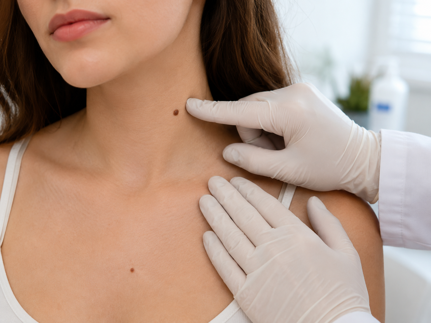
This guide explains what a seborrheic keratosis is, the visual features that distinguish it from a mole, and the situations where it still earns a 30-minute consultation. Written by a GMC-registered consultant plastic surgeon for adults across the UK.
What a seborrheic keratosis actually is
A seborrheic keratosis (plural: keratoses; abbreviated SK) is a benign overgrowth of keratinocytes, the cells that form the visible surface of the skin. They are not made of melanocytes, which is what distinguishes them biologically from moles. The pigment in an SK comes from melanin trapped in the thickened keratinocyte layer rather than from a melanocytic cluster, which is why SKs can appear dark without the underlying biology of a true pigmented lesion.[1]
The cause is not fully understood. Genetic predisposition plays a role (SKs run in families), age plays a strong role (they're rare under 40, common at 50+, very common at 70+), and chronic UV exposure may contribute, but they appear on covered skin too. Most adults over 60 have several; many over 70 have dozens. They never become malignant; SKs do not progress to melanoma, basal cell carcinoma, or any other cancer.
What's important: an SK and a melanoma can superficially resemble each other, particularly when the SK is dark, irregular, or recently inflamed. The clinical mistake patients make most often is dismissing an actual melanoma as 'probably another one of those harmless thingies'. Dermatoscopy distinguishes them reliably; self-assessment alone does not.
The 'stuck-on' appearance
The classical clinical description of a seborrheic keratosis is stuck-on: it looks as if a piece of brown wax or putty has been pressed onto the surface of the skin and could be peeled off. The edge is sharp, often with a slightly raised border that's clearly demarcated from surrounding normal skin. The surface is typically rough, warty, sometimes scaly, often with small pinpoint dark dots (horn cysts) visible on close inspection.
By contrast, a mole tends to look integrated with the surrounding skin: the pigment is in the skin, not on it, and the border, while clear, doesn't have the sharp peelable quality of an SK. Mole surfaces are typically smooth or very slightly textured (cobblestone for compound naevi); SK surfaces are rougher, more uneven, often with a slight greasy or wax-like feel under the fingertip.
Practical home test. Run your fingertip lightly across the lesion. Mole: smooth or very slightly textured, feels like 'just skin with pigment'. SK: rough, slightly raised border, sometimes greasy or warty texture, feels like a small piece of something stuck on. The test isn't perfect (some SKs are flat, some intradermal naevi are dome-shaped) but it's right in the great majority of cases for adults over 50.
Common SK presentations across the body
Trunk: the chest, back and abdomen are the most common sites. SKs here are typically tan to dark brown, 5-15mm, slightly raised, with the classical stuck-on appearance. Many adults over 60 have ten or more on the back alone, often noticed when getting changed or by a partner.
Face and temples: SKs on the face tend to be smaller and flatter than on the trunk, sometimes blending into the surrounding skin texture. Around the temples and hairline they can develop a 'dermatosis papulosa nigra' pattern, multiple small dark papules clustered together, more common in patients with skin types V-VI.
Sun-exposed sites in older adults: SKs on the dorsum of the hands, forearms and lower legs are typical of patients over 70. They can be quite warty and irregular and are sometimes confused with squamous cell carcinoma or with lentigo maligna. This is where the dermatoscopic distinction becomes most important; the visual appearance overlaps meaningfully with concerning lesions.
When an SK still warrants a consultant
Three scenarios where what looks like an SK still needs a consultant's eye. The lesion is changing. SKs are usually stable across years; if a 'stuck-on' lesion is visibly growing, darkening, or developing irregular features over months, treat it as an unidentified pigmented lesion rather than an assumed SK and book the consultation.
The lesion is irritated, bleeding or crusting. Inflamed SKs (irritated by friction, scratching, or trauma) can bleed and crust, which is the same warning sign for melanoma and other skin cancers. The pattern matters: an SK that bleeds once after physical trauma and heals cleanly is benign; an SK that bleeds spontaneously, crusts repeatedly, or fails to heal after irritation deserves dermatoscopy.
You can't confidently identify it. If the lesion looks 'half mole, half SK', has features of both, or sits at the boundary of your visual confidence, don't try to talk yourself into one or the other. Book the consultation; £250 buys you dermatoscopic certainty in 30 minutes, and the cost of being wrong about a melanoma masquerading as an SK is much higher than the cost of an unnecessary appointment.
Removal: when, how, and at what cost
Most SKs do not need removal. They are entirely benign and don't progress to anything. Patients ask about removal for one of three reasons: cosmetic appearance (an SK on the face that bothers them), repeated irritation (an SK that catches on bra straps, collars, or jewellery), or diagnostic uncertainty (a lesion the consultant wants to remove and send to histology to be definitively certain it isn't melanoma).
Cosmetic SK removal is straightforward. Three techniques are commonly used: shave excision (a blade trims the lesion flush with surrounding skin, with a flat scar that usually heals invisibly), electrocautery (precision heat ablation, suitable for small lesions, fast healing), and cryotherapy (liquid nitrogen freezes the lesion which then sloughs off over a week or two, sometimes leaving a small pale patch). All three are appropriate for confirmed-benign SKs; none destroys diagnostic information meaningfully because SKs don't have the histology stakes a true mole has.
At our clinic, SK removal is usually shave excision or electrocautery under local anaesthetic. Pricing follows our standard small-lesion structure: £275 first lesion, £175 each additional, no separate histology fee unless the consultant decides to send it (in which case £85-150 lab fee applies). Most patients with multiple SKs have several removed in a single appointment.
What the consultant will do at the appointment
If you book a mole check or skin cancer check for what you suspect is an SK but want assessed, the appointment runs the standard 30-minute structure: clinical history, naked-eye examination, dermatoscopy of every lesion of concern. Dermatoscopy reveals the specific features of an SK (milia-like cysts, comedo-like openings, fissures and ridges) that distinguish it from a melanocytic lesion at high reliability.[2]
If the lesion is dermatoscopically a confirmed SK, the consultant will reassure you and discuss whether removal is wanted (cosmetic, irritation-driven, or not at all). If it's dermatoscopically borderline, excision and histology is the safer route. If it's dermatoscopically suspicious for melanoma despite the SK-like surface, full-thickness excision is recommended same-day.
Most patients in this position leave with reassurance about the specific lesion they were worried about, a brief tour of any other SKs the consultant noticed in passing, and (often) a quote for cosmetic removal of one or two if they want it. The £250 fee covers the consultation in full; removal is billed separately if you proceed.
What not to do at home
Three home interventions to avoid. Do not pick or scratch a suspected SK. It can become inflamed, bleed, and crust, all of which mimic the warning signs of melanoma and complicate a future consultant assessment. Inflamed SKs are also harder to dermatoscope cleanly, so you may end up with an inconclusive examination that requires re-imaging in a few weeks.
Do not use over-the-counter mole, wart or 'skin tag' removal kits on a suspected SK. They contain caustic agents (salicylic acid, trichloroacetic acid) designed for non-pigmented lesions. Applied to an SK they damage the surface, distort the dermatoscopic appearance, and can mask any underlying features the consultant would otherwise have used to confirm benignity. They also cannot 'remove' an SK reliably; they only damage the surface.
Do not assume any new dark lesion is just another SK without checking. This is the mistake we see most often: patients accumulate SKs across decades, get used to ignoring them, and later miss a new pigmented lesion that's actually a melanoma because they assumed it was 'one of those harmless thingies'. Trust dermatoscopy, not the assumption.
Common questions
Frequently asked
References
Sources cited
- Hafner C, Vogt T. Seborrheic keratosis. J Dtsch Dermatol Ges. 2008;6(8):664-677. View source
- Braun RP, Rabinovitz HS, Krischer J, et al. Dermoscopy of pigmented seborrheic keratosis: a morphological study. Arch Dermatol. 2002;138(12):1556-1560. View source
- Pierard GE, Pierard-Franchimont C, Laso Dosal F, et al. Pigmentary changes in skin senescence. J Eur Acad Dermatol Venereol. 1995;5(3):222-228.
- British Association of Dermatologists. Patient information leaflet on seborrhoeic keratoses. View source
- Roh NK, Hahn HJ, Lee YW, et al. Clinical and histopathological features of skin lesions on the face: a review of 1,250 cases. Ann Dermatol. 2016;28(2):152-158. View source